 Add to Chrome
 Add to Firefox
 Add to Edge
Do 3D Medical Foundation Models See Through MRI Artifacts? A Controlled Study of Representation Robustness
Aug 06, 2026
Self-supervised 3D medical foundation models are increasingly used as general-purpose feature extractors, yet their sensitivity to MRI artifacts remains poorly understood. We present a controlled evaluation of representation robustness across five pretrained 3D encoders spanning different architectures, objectives, pretraining domains, and dataset scales. Using BraTS-Africa cases with four MRI sequences, we generate seven frequency- and image-domain artifacts at five predefined corruption settings. Robustness is assessed using linear centered kernel alignment (CKA), RankMe, and UMAP, complemented by an independent segmentation-consistency analysis. We find that robustness is strongly model- and artifact-dependent. 3DINO exhibits the most consistently stable representations, while BrainIAC is highly sensitive to several corruptions; NeuroVFM, BrainFM, and Neuro-SimCLR show intermediate but distinct artifact-specific profiles. Across many conditions, CKA decreases substantially while RankMe remains comparatively stable, indicating that artifacts often distort representation geometry without causing dimensional collapse. Segmentation consistency also degrades under corruption, particularly for ghosting and Rician noise, but aligns only partially with representation-level robustness. These findings show that larger-scale or domain-specific pretraining alone does not guarantee artifact invariance and motivate explicit robustness evaluation before deploying 3D foundation models in heterogeneous MRI settings.

Validation of Diagnostic Artificial Intelligence Models for Prostate Pathology in a Middle Eastern Cohort
Dec 19, 2025Background: Artificial intelligence (AI) is improving the efficiency and accuracy of cancer diagnostics. The performance of pathology AI systems has been almost exclusively evaluated on European and US cohorts from large centers. For global AI adoption in pathology, validation studies on currently under-represented populations - where the potential gains from AI support may also be greatest - are needed. We present the first study with an external validation cohort from the Middle East, focusing on AI-based diagnosis and Gleason grading of prostate cancer. Methods: We collected and digitised 339 prostate biopsy specimens from the Kurdistan region, Iraq, representing a consecutive series of 185 patients spanning the period 2013-2024. We evaluated a task-specific end-to-end AI model and two foundation models in terms of their concordance with pathologists and consistency across samples digitised on three scanner models (Hamamatsu, Leica, and Grundium). Findings: Grading concordance between AI and pathologists was similar to pathologist-pathologist concordance with Cohen's quadratically weighted kappa 0.801 vs. 0.799 (p=0.9824). Cross-scanner concordance was high (quadratically weighted kappa > 0.90) for all AI models and scanner pairs, including low-cost compact scanner. Interpretation: AI models demonstrated pathologist-level performance in prostate histopathology assessment. Compact scanners can provide a route for validation studies in non-digitalised settings and enable cost-effective adoption of AI in laboratories with limited sample volumes. This first openly available digital pathology dataset from the Middle East supports further research into globally equitable AI pathology. Funding: SciLifeLab and Wallenberg Data Driven Life Science Program, Instrumentarium Science Foundation, Karolinska Institutet Research Foundation.
Foundation Models -- A Panacea for Artificial Intelligence in Pathology?
Feb 28, 2025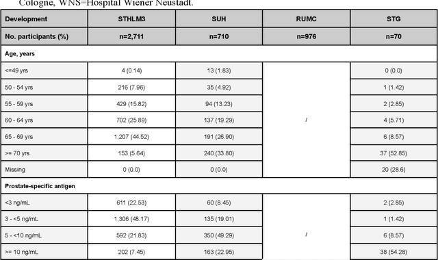
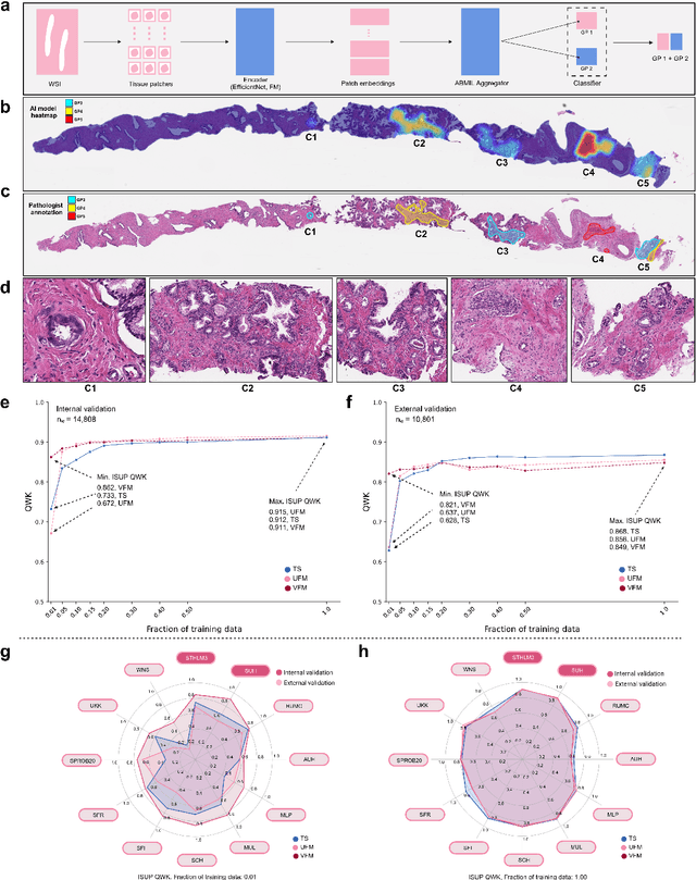
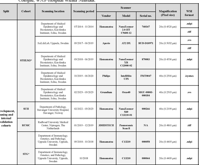
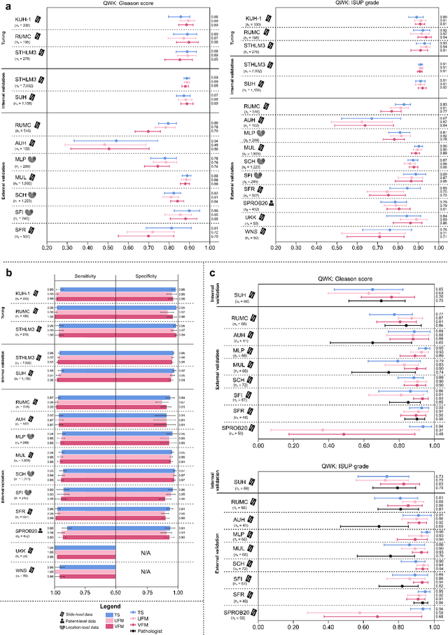
The role of artificial intelligence (AI) in pathology has evolved from aiding diagnostics to uncovering predictive morphological patterns in whole slide images (WSIs). Recently, foundation models (FMs) leveraging self-supervised pre-training have been widely advocated as a universal solution for diverse downstream tasks. However, open questions remain about their clinical applicability and generalization advantages over end-to-end learning using task-specific (TS) models. Here, we focused on AI with clinical-grade performance for prostate cancer diagnosis and Gleason grading. We present the largest validation of AI for this task, using over 100,000 core needle biopsies from 7,342 patients across 15 sites in 11 countries. We compared two FMs with a fully end-to-end TS model in a multiple instance learning framework. Our findings challenge assumptions that FMs universally outperform TS models. While FMs demonstrated utility in data-scarce scenarios, their performance converged with - and was in some cases surpassed by - TS models when sufficient labeled training data were available. Notably, extensive task-specific training markedly reduced clinically significant misgrading, misdiagnosis of challenging morphologies, and variability across different WSI scanners. Additionally, FMs used up to 35 times more energy than the TS model, raising concerns about their sustainability. Our results underscore that while FMs offer clear advantages for rapid prototyping and research, their role as a universal solution for clinically applicable medical AI remains uncertain. For high-stakes clinical applications, rigorous validation and consideration of task-specific training remain critically important. We advocate for integrating the strengths of FMs and end-to-end learning to achieve robust and resource-efficient AI pathology solutions fit for clinical use.