 Add to Chrome
 Add to Firefox
 Add to Edge
Deep learning classifier of locally advanced rectal cancer treatment response from endoscopy images
May 06, 2024


We developed a deep learning classifier of rectal cancer response (tumor vs. no-tumor) to total neoadjuvant treatment (TNT) from endoscopic images acquired before, during, and following TNT. We further evaluated the network's ability in a near out-of-distribution (OOD) problem to identify local regrowth (LR) from follow-up endoscopy images acquired several months to years after completing TNT. We addressed endoscopic image variability by using optimal mass transport-based image harmonization. We evaluated multiple training regularization schemes to study the ResNet-50 network's in-distribution and near-OOD generalization ability. Test time augmentation resulted in the most considerable accuracy improvement. Image harmonization resulted in slight accuracy improvement for the near-OOD cases. Our results suggest that off-the-shelf deep learning classifiers can detect rectal cancer from endoscopic images at various stages of therapy for surveillance.
Trustworthiness of Pretrained Transformers for Lung Cancer Segmentation
Mar 19, 2024We assessed the trustworthiness of two self-supervision pretrained transformer models, Swin UNETR and SMIT, for fine-tuned lung (LC) tumor segmentation using 670 CT and MRI scans. We measured segmentation accuracy on two public 3D-CT datasets, robustness on CT scans of patients with COVID-19, CT scans of patients with ovarian cancer and T2-weighted MRI of men with prostate cancer, and zero-shot generalization of LC for T2-weighted MRIs. Both models demonstrated high accuracy on in-distribution data (Dice 0.80 for SMIT and 0.78 for Swin UNETR). SMIT showed similar near-out-of-distribution performance on CT scans (AUROC 89.85% vs. 89.19%) but significantly better far-out-of-distribution accuracy on CT (AUROC 97.2% vs. 87.1%) and MRI (92.15% vs. 73.8%). SMIT outperformed Swin UNETR in zero-shot segmentation on MRI (Dice 0.78 vs. 0.69). We expect these findings to guide the safe development and deployment of current and future pretrained models in routine clinical use.
Self-distilled Masked Attention guided masked image modeling with noise Regularized Teacher (SMART) for medical image analysis
Oct 02, 2023


Hierarchical shifted window transformers (Swin) are a computationally efficient and more accurate alternative to plain vision transformers. Masked image modeling (MIM)-based pretraining is highly effective in increasing models' transferability to a variety of downstream tasks. However, more accurate and efficient attention guided MIM approaches are difficult to implement with Swin due to it's lack of an explicit global attention. We thus architecturally enhanced Swin with semantic class attention for self-supervised attention guided co-distillation with MIM. We also introduced a noise injected momentum teacher, implemented with patch dropout of teacher's inputs for improved training regularization and accuracy. Our approach, called \underline{s}elf-distilled \underline{m}asked \underline{a}ttention MIM with noise \underline{r}egularized \underline{t}eacher (SMART) was pretrained with \textbf{10,412} unlabeled 3D computed tomography (CT)s of multiple disease sites and sourced from institutional and public datasets. We evaluated SMART for multiple downstream tasks involving analysis of 3D CTs of lung cancer (LC) patients for: (i) [Task I] predicting immunotherapy response in advanced stage LC (n = 200 internal dataset), (ii) [Task II] predicting LC recurrence in early stage LC before surgery (n = 156 public dataset), (iii) [Task III] LC segmentation (n = 200 internal, 21 public dataset), and (iv) [Task IV] unsupervised clustering of organs in the chest and abdomen (n = 1,743 public dataset) \underline{without} finetuning. SMART predicted immunotherapy response with an AUC of 0.916, LC recurrence with an AUC of 0.793, segmented LC with Dice accuracy of 0.81, and clustered organs with an inter-class cluster distance of 5.94, indicating capability of attention guided MIM for Swin in medical image analysis.
Deep Learning Based Dominant Index Lesion Segmentation for MR-guided Radiation Therapy of Prostate Cancer
Mar 06, 2023Dose escalation radiotherapy allows increased control of prostate cancer (PCa) but requires segmentation of dominant index lesions (DIL), motivating the development of automated methods for fast, accurate, and consistent segmentation of PCa DIL. We evaluated five deep-learning networks on apparent diffusion coefficient (ADC) MRI from 500 lesions in 365 patients arising from internal training Dataset 1 (1.5Tesla GE MR with endorectal coil), external ProstateX Dataset 2 (3Tesla Siemens MR), and internal inter-rater Dataset 3 (3Tesla Philips MR). The networks include: multiple resolution residually connected network (MRRN) and MRRN regularized in training with deep supervision (MRRN-DS), Unet, Unet++, ResUnet, and fast panoptic segmentation (FPSnet) as well as fast panoptic segmentation with smoothed labels (FPSnet-SL). Models were evaluated by volumetric DIL segmentation accuracy using Dice similarity coefficient (DSC) and detection accuracy, as a function of lesion aggressiveness, size, and location (Dataset 1 and 2), and accuracy with respect to two-raters (on Dataset 3). In general MRRN-DS more accurately segmented tumors than other methods on the testing datasets. MRRN-DS significantly outperformed ResUnet in Dataset2 (DSC of 0.54 vs. 0.44, p<0.001) and the Unet++ in Dataset3 (DSC of 0.45 vs. p=0.04). FPSnet-SL was similarly accurate as MRRN-DS in Dataset2 (p = 0.30), but MRRN-DS significantly outperformed FPSnet and FPSnet-SL in both Dataset1 (0.60 vs 0.51 [p=0.01] and 0.54 [p=0.049] respectively) and Dataset3 (0.45 vs 0.06 [p=0.002] and 0.24 [p=0.004] respectively). Finally, MRRN-DS produced slightly higher agreement with experienced radiologist than two radiologists in Dataset 3 (DSC of 0.45 vs. 0.41).
* https://pubmed.ncbi.nlm.nih.gov/36856092/
Progressively refined deep joint registration segmentation (ProRSeg) of gastrointestinal organs at risk: Application to MRI and cone-beam CT
Oct 25, 2022


Method: ProRSeg was trained using 5-fold cross-validation with 110 T2-weighted MRI acquired at 5 treatment fractions from 10 different patients, taking care that same patient scans were not placed in training and testing folds. Segmentation accuracy was measured using Dice similarity coefficient (DSC) and Hausdorff distance at 95th percentile (HD95). Registration consistency was measured using coefficient of variation (CV) in displacement of OARs. Ablation tests and accuracy comparisons against multiple methods were done. Finally, applicability of ProRSeg to segment cone-beam CT (CBCT) scans was evaluated on 80 scans using 5-fold cross-validation. Results: ProRSeg processed 3D volumes (128 $\times$ 192 $\times$ 128) in 3 secs on a NVIDIA Tesla V100 GPU. It's segmentations were significantly more accurate ($p<0.001$) than compared methods, achieving a DSC of 0.94 $\pm$0.02 for liver, 0.88$\pm$0.04 for large bowel, 0.78$\pm$0.03 for small bowel and 0.82$\pm$0.04 for stomach-duodenum from MRI. ProRSeg achieved a DSC of 0.72$\pm$0.01 for small bowel and 0.76$\pm$0.03 for stomach-duodenum from CBCT. ProRSeg registrations resulted in the lowest CV in displacement (stomach-duodenum $CV_{x}$: 0.75\%, $CV_{y}$: 0.73\%, and $CV_{z}$: 0.81\%; small bowel $CV_{x}$: 0.80\%, $CV_{y}$: 0.80\%, and $CV_{z}$: 0.68\%; large bowel $CV_{x}$: 0.71\%, $CV_{y}$ : 0.81\%, and $CV_{z}$: 0.75\%). ProRSeg based dose accumulation accounting for intra-fraction (pre-treatment to post-treatment MRI scan) and inter-fraction motion showed that the organ dose constraints were violated in 4 patients for stomach-duodenum and for 3 patients for small bowel. Study limitations include lack of independent testing and ground truth phantom datasets to measure dose accumulation accuracy.
Self-supervised 3D anatomy segmentation using self-distilled masked image transformer (SMIT)
May 20, 2022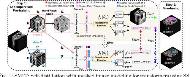

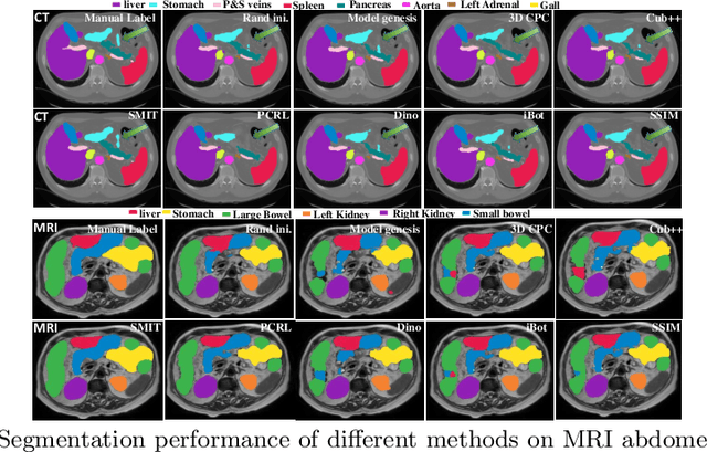
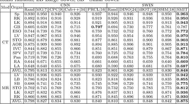
Vision transformers, with their ability to more efficiently model long-range context, have demonstrated impressive accuracy gains in several computer vision and medical image analysis tasks including segmentation. However, such methods need large labeled datasets for training, which is hard to obtain for medical image analysis. Self-supervised learning (SSL) has demonstrated success in medical image segmentation using convolutional networks. In this work, we developed a \underline{s}elf-distillation learning with \underline{m}asked \underline{i}mage modeling method to perform SSL for vision \underline{t}ransformers (SMIT) applied to 3D multi-organ segmentation from CT and MRI. Our contribution is a dense pixel-wise regression within masked patches called masked image prediction, which we combined with masked patch token distillation as pretext task to pre-train vision transformers. We show our approach is more accurate and requires fewer fine tuning datasets than other pretext tasks. Unlike prior medical image methods, which typically used image sets arising from disease sites and imaging modalities corresponding to the target tasks, we used 3,643 CT scans (602,708 images) arising from head and neck, lung, and kidney cancers as well as COVID-19 for pre-training and applied it to abdominal organs segmentation from MRI pancreatic cancer patients as well as publicly available 13 different abdominal organs segmentation from CT. Our method showed clear accuracy improvement (average DSC of 0.875 from MRI and 0.878 from CT) with reduced requirement for fine-tuning datasets over commonly used pretext tasks. Extensive comparisons against multiple current SSL methods were done. Code will be made available upon acceptance for publication.
Wasserstein Image Local Analysis: Histogram of Orientations, Smoothing and Edge Detection
May 11, 2022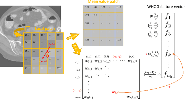
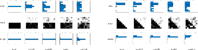
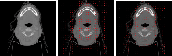
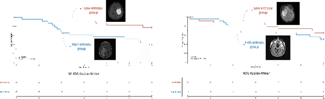
The Histogram of Oriented Gradient is a widely used image feature, which describes local image directionality based on numerical differentiation. Due to its ill-posed nature, small noise may lead to large errors. Conventional HOG may fail to produce meaningful directionality results in the presence of noise, which is common in medical radiographic imaging. We approach the directionality problem from a novel perspective by the use of the optimal transport map of a local image patch to a uni-color patch of its mean. We decompose the transport map into sub-work costs in different directions. We evaluated the ability of the optimal transport to quantify tumor heterogeneity from brain MRI images of patients with glioblastoma multiforme from the TCIA. By considering the entropy difference of the extracted local directionality within tumor regions, we found that patients with higher entropy in their images, had statistically significant worse overall survival (p $=0.008$), which indicates that tumors exhibiting flows in many directions may be more malignant, perhaps reflecting high tumor histologic grade, a reflection of histologic disorganization. We also explored the possibility of solving classical image processing problems such as smoothing and edge detection via optimal transport. By looking for a 2-color patch with minimum transport distance to a local patch, we derive a nonlinear shock filter, which preserves edges. Moreover, we found that the color difference of the computed 2-color patch indicates whether there is a large change in color, i.e., an edge in the given patch. In summary, we expand the usefulness of optimal transport as an image local analysis tool, to extract robust measures of imaging tumor heterogeneity for outcomes prediction as well as image pre-processing. Because of its robust nature, we find it offers several advantages over the classical approaches.
One shot PACS: Patient specific Anatomic Context and Shape prior aware recurrent registration-segmentation of longitudinal thoracic cone beam CTs
Jan 26, 2022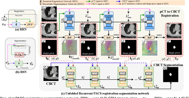
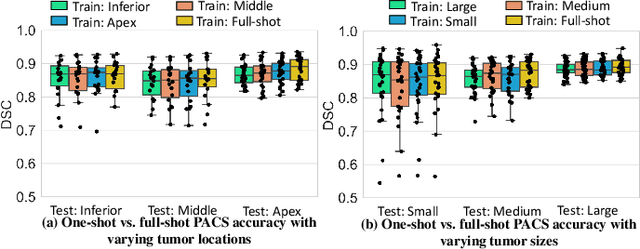
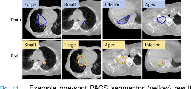
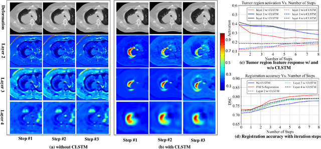
Image-guided adaptive lung radiotherapy requires accurate tumor and organs segmentation from during treatment cone-beam CT (CBCT) images. Thoracic CBCTs are hard to segment because of low soft-tissue contrast, imaging artifacts, respiratory motion, and large treatment induced intra-thoracic anatomic changes. Hence, we developed a novel Patient-specific Anatomic Context and Shape prior or PACS-aware 3D recurrent registration-segmentation network for longitudinal thoracic CBCT segmentation. Segmentation and registration networks were concurrently trained in an end-to-end framework and implemented with convolutional long-short term memory models. The registration network was trained in an unsupervised manner using pairs of planning CT (pCT) and CBCT images and produced a progressively deformed sequence of images. The segmentation network was optimized in a one-shot setting by combining progressively deformed pCT (anatomic context) and pCT delineations (shape context) with CBCT images. Our method, one-shot PACS was significantly more accurate (p$<$0.001) for tumor (DSC of 0.83 $\pm$ 0.08, surface DSC [sDSC] of 0.97 $\pm$ 0.06, and Hausdorff distance at $95^{th}$ percentile [HD95] of 3.97$\pm$3.02mm) and the esophagus (DSC of 0.78 $\pm$ 0.13, sDSC of 0.90$\pm$0.14, HD95 of 3.22$\pm$2.02) segmentation than multiple methods. Ablation tests and comparative experiments were also done.
Unpaired cross-modality educed distillation (CMEDL) applied to CT lung tumor segmentation
Jul 16, 2021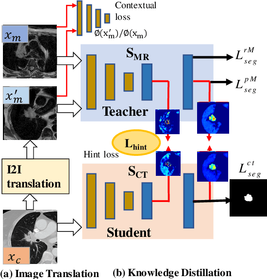
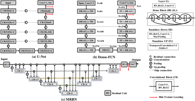
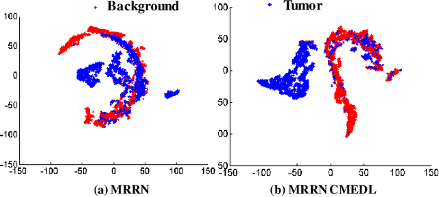
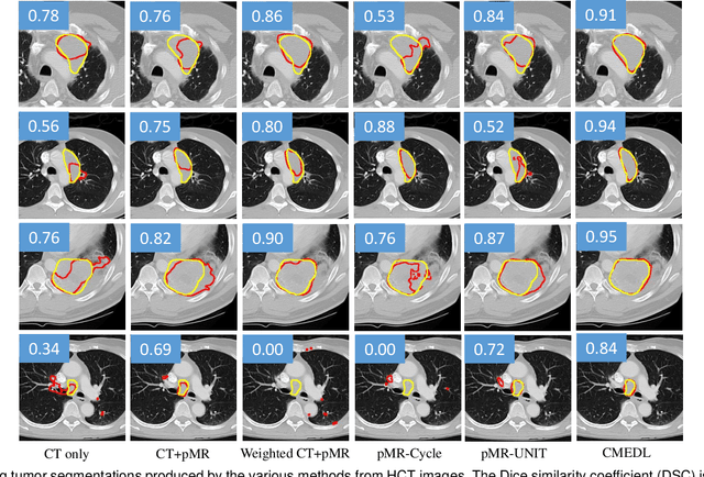
Accurate and robust segmentation of lung cancers from CTs is needed to more accurately plan and deliver radiotherapy and to measure treatment response. This is particularly difficult for tumors located close to mediastium, due to low soft-tissue contrast. Therefore, we developed a new cross-modality educed distillation (CMEDL) approach, using unpaired CT and MRI scans, whereby a teacher MRI network guides a student CT network to extract features that signal the difference between foreground and background. Our contribution eliminates two requirements of distillation methods: (i) paired image sets by using an image to image (I2I) translation and (ii) pre-training of the teacher network with a large training set by using concurrent training of all networks. Our framework uses an end-to-end trained unpaired I2I translation, teacher, and student segmentation networks. Our framework can be combined with any I2I and segmentation network. We demonstrate our framework's feasibility using 3 segmentation and 2 I2I methods. All networks were trained with 377 CT and 82 T2w MRI from different sets of patients. Ablation tests and different strategies for incorporating MRI information into CT were performed. Accuracy was measured using Dice similarity (DSC), surface Dice (sDSC), and Hausdorff distance at the 95$^{th}$ percentile (HD95). The CMEDL approach was significantly (p $<$ 0.001) more accurate than non-CMEDL methods, quantitatively and visually. It produced the highest segmentation accuracy (sDSC of 0.83 $\pm$ 0.16 and HD95 of 5.20 $\pm$ 6.86mm). CMEDL was also more accurate than using either pMRI's or the combination of CT's with pMRI's for segmentation.
Deep cross-modality (MR-CT) educed distillation learning for cone beam CT lung tumor segmentation
Feb 26, 2021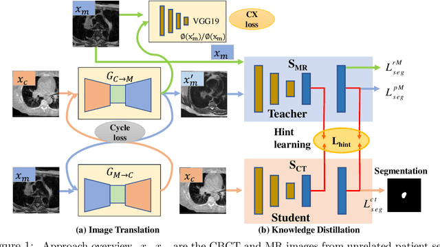

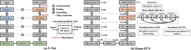
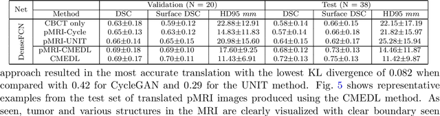
Despite the widespread availability of in-treatment room cone beam computed tomography (CBCT) imaging, due to the lack of reliable segmentation methods, CBCT is only used for gross set up corrections in lung radiotherapies. Accurate and reliable auto-segmentation tools could potentiate volumetric response assessment and geometry-guided adaptive radiation therapies. Therefore, we developed a new deep learning CBCT lung tumor segmentation method. Methods: The key idea of our approach called cross modality educed distillation (CMEDL) is to use magnetic resonance imaging (MRI) to guide a CBCT segmentation network training to extract more informative features during training. We accomplish this by training an end-to-end network comprised of unpaired domain adaptation (UDA) and cross-domain segmentation distillation networks (SDN) using unpaired CBCT and MRI datasets. Feature distillation regularizes the student network to extract CBCT features that match the statistical distribution of MRI features extracted by the teacher network and obtain better differentiation of tumor from background.} We also compared against an alternative framework that used UDA with MR segmentation network, whereby segmentation was done on the synthesized pseudo MRI representation. All networks were trained with 216 weekly CBCTs and 82 T2-weighted turbo spin echo MRI acquired from different patient cohorts. Validation was done on 20 weekly CBCTs from patients not used in training. Independent testing was done on 38 weekly CBCTs from patients not used in training or validation. Segmentation accuracy was measured using surface Dice similarity coefficient (SDSC) and Hausdroff distance at 95th percentile (HD95) metrics.