 Add to Chrome
 Add to Firefox
 Add to Edge
Transfer learning for diagnosis of congenital abnormalities of the kidney and urinary tract in children based on Ultrasound imaging data
Dec 31, 2017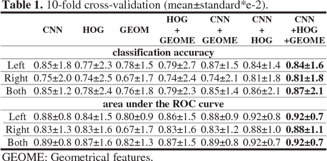
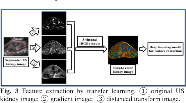
Classification of ultrasound (US) kidney images for diagnosis of congenital abnormalities of the kidney and urinary tract (CAKUT) in children is a challenging task. It is desirable to improve existing pattern classification models that are built upon conventional image features. In this study, we propose a transfer learning-based method to extract imaging features from US kidney images in order to improve the CAKUT diagnosis in children. Particularly, a pre-trained deep learning model (imagenet-caffe-alex) is adopted for transfer learning-based feature extraction from 3-channel feature maps computed from US images, including original images, gradient features, and distanced transform features. Support vector machine classifiers are then built upon different sets of features, including the transfer learning features, conventional imaging features, and their combination. Experimental results have demonstrated that the combination of transfer learning features and conventional imaging features yielded the best classification performance for distinguishing CAKUT patients from normal controls based on their US kidney images.
A dynamic graph-cuts method with integrated multiple feature maps for segmenting kidneys in ultrasound images
Jun 11, 2017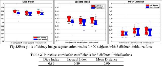
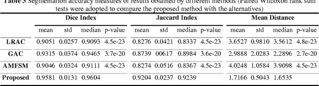
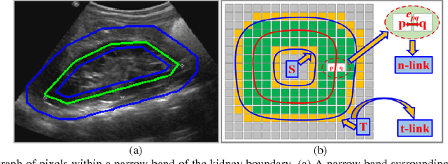
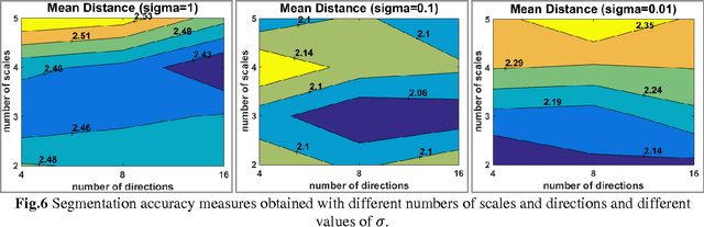
Purpose: To improve kidney segmentation in clinical ultrasound (US) images, we develop a new graph cuts based method to segment kidney US images by integrating original image intensity information and texture feature maps extracted using Gabor filters. Methods: To handle large appearance variation within kidney images and improve computational efficiency, we build a graph of image pixels close to kidney boundary instead of building a graph of the whole image. To make the kidney segmentation robust to weak boundaries, we adopt localized regional information to measure similarity between image pixels for computing edge weights to build the graph of image pixels. The localized graph is dynamically updated and the GC based segmentation iteratively progresses until convergence. The proposed method has been evaluated and compared with state of the art image segmentation methods based on clinical kidney US images of 85 subjects. We randomly selected US images of 20 subjects as training data for tuning the parameters, and validated the methods based on US images of the remaining 65 subjects. The segmentation results have been quantitatively analyzed using 3 metrics, including Dice Index, Jaccard Index, and Mean Distance. Results: Experiment results demonstrated that the proposed method obtained segmentation results for bilateral kidneys of 65 subjects with average Dice index of 0.9581, Jaccard index of 0.9204, and Mean Distance of 1.7166, better than other methods under comparison (p<10-19, paired Wilcoxon rank sum tests). Conclusions: The proposed method achieved promising performance for segmenting kidneys in US images, better than segmentation methods that built on any single channel of image information. This method will facilitate extraction of kidney characteristics that may predict important clinical outcomes such progression chronic kidney disease.