 Add to Chrome
 Add to Firefox
 Add to Edge
An Agentic AI Framework Overcomes Fundamental Limitations of Large Language Models for Glaucoma Detection from Fundus Photography
Aug 07, 2026
Large language models (LLMs) show promise in medical image interpretation but suffer from hallucination, limited accuracy, and run-to-run inconsistency. We developed and validated an agentic AI framework integrating LLMs with specialized deep learning tools for glaucoma detection from fundus photography. The workflow had three steps: (1) LLM initial assessment; (2) function calling to invoke specialized tools for image quality (QAModel, FundaQ-8), glaucoma classification (SwinV2-Tiny), and optic disc/cup segmentation (SegFormer-B0); and (3) LLM reflection integrating the initial impression with tool outputs. Two LLMs (Gemini 2.5 Flash, GPT-5.4 mini) were evaluated on two public datasets (ORIGA, n=100; RIM-ONE-v3, n=100) under uncropped and cropped fields of view; all images were independently graded by a masked fellowship-trained glaucoma specialist. The agentic workflow improved classification accuracy by 16 to 47 percentage points across all conditions, reaching within 6 points of the specialist; on RIM-ONE-v3 the best configurations matched the specialist accuracy of 88%. LLM-alone approaches failed in two ways: GPT-5.4 mini showed positive bias (sensitivity 95-100%, specificity 0-5%), while Gemini 2.5 Flash varied stochastically between runs; the agentic workflow corrected both. Cup-to-disc ratio error fell 15-50% (MAE 0.156-0.228 to 0.104-0.132), and correlation with specialist grading rose from weak (r=0.12-0.39) to moderate-strong (r=0.59-0.84). Run-to-run consistency rose from near-random (kappa as low as -0.01) to near-perfect (kappa up to 0.96). Integrating LLMs with specialized tools addressed key limitations of LLM-alone approaches, including over-diagnosis and run-to-run variability. Gains held for both LLMs, suggesting generalizability across backbones, and may signal a shift from monolithic models toward orchestrated multi-agent systems in medical AI.

One-Vote Veto: A Self-Training Strategy for Low-Shot Learning of a Task-Invariant Embedding to Diagnose Glaucoma
Jan 19, 2021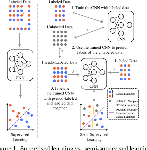

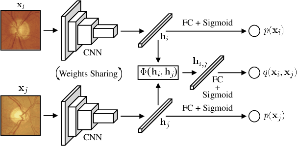
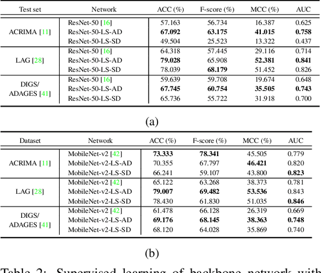
Convolutional neural networks (CNNs) are a promising technique for automated glaucoma diagnosis from images of the fundus, and these images are routinely acquired as part of an ophthalmic exam. Nevertheless, CNNs typically require a large amount of well-labeled data for training, which may not be available in many biomedical image classification applications, especially when diseases are rare and where labeling by experts is costly. This paper makes two contributions to address this issue: (1) It introduces a new network architecture and training method for low-shot learning when labeled data are limited and imbalanced, and (2) it introduces a new semi-supervised learning strategy that uses additional unlabeled training data to achieve great accuracy. Our multi-task twin neural network (MTTNN) can use any backbone CNN, and we demonstrate with ResNet-50 and MobileNet-v2 that its accuracy with limited training data approaches the accuracy of a finetuned backbone trained with a dataset that is 50 times larger. We also introduce One-Vote Veto (OVV) self-training, a semi-supervised learning strategy, that is designed specifically for MTTNNs. By taking both self-predictions and contrastive-predictions of the unlabeled training data into account, OVV self-training provides additional pseudo labels for finetuning a pretrained MTTNN. Using a large dataset with more than 50,000 fundus images acquired over 25 years, extensive experimental results demonstrate the effectiveness of low-shot learning with MTTNN and semi-supervised learning with OVV. Three additional, smaller clinical datasets of fundus images acquired under different conditions (cameras, instruments, locations, populations), are used to demonstrate generalizability of the methods. Source code and pretrained models will be publicly available upon publication.