 Add to Chrome
 Add to Firefox
 Add to Edge
Swap-Free Fat-Water Separation in Dixon MRI using Conditional Generative Adversarial Networks
Jul 29, 2021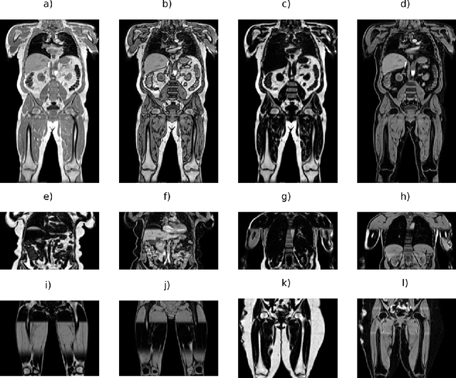
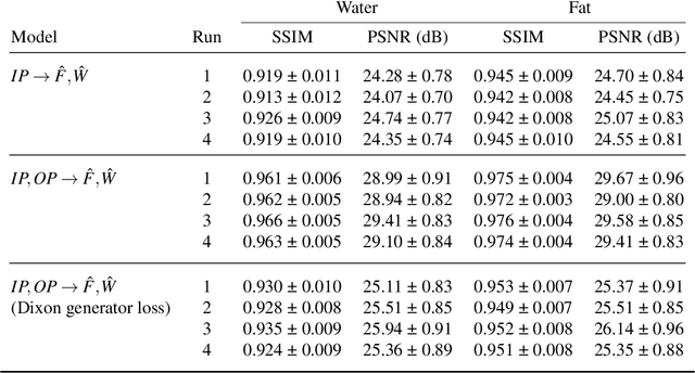
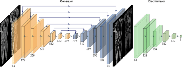
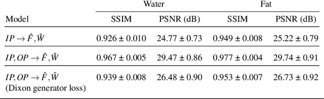
Dixon MRI is widely used for body composition studies. Current processing methods associated with large whole-body volumes are time intensive and prone to artifacts during fat-water separation performed on the scanner, making the data difficult to analyse. The most common artifact are fat-water swaps, where the labels are inverted at the voxel level. It is common for researchers to discard swapped data (generally around 10%), which can be wasteful and lead to unintended biases. The UK Biobank is acquiring Dixon MRI for over 100,000 participants, and thousands of swaps will occur. If those go undetected, errors will propagate into processes such as abdominal organ segmentation and dilute the results in population-based analyses. There is a clear need for a fast and robust method to accurately separate fat and water channels. In this work we propose such a method based on style transfer using a conditional generative adversarial network. We also introduce a new Dixon loss function for the generator model. Using data from the UK Biobank Dixon MRI, our model is able to predict highly accurate fat and water channels that are free from artifacts. We show that the model separates fat and water channels using either single input (in-phase) or dual input (in-phase and opposed-phase), with the latter producing improved results. Our proposed method enables faster and more accurate downstream analysis of body composition from Dixon MRI in population studies by eliminating the need for visual inspection or discarding data due to fat-water swaps.
Large-Scale Analysis of Iliopsoas Muscle Volumes in the UK Biobank
Aug 14, 2020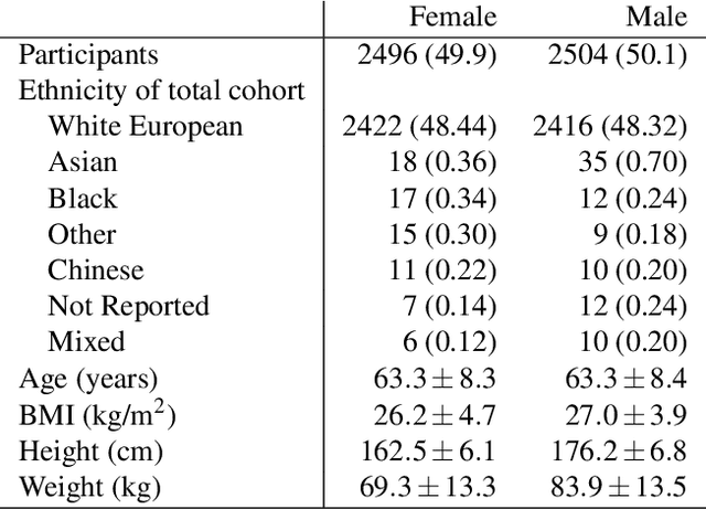
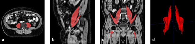
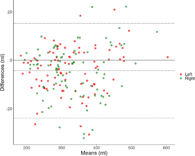
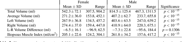
Psoas muscle measurements are frequently used as markers of sarcopenia and predictors of health. Manually measured cross-sectional areas are most commonly used, but there is a lack of consistency regarding the position of the measurementand manual annotations are not practical for large population studies. We have developed a fully automated method to measure iliopsoas muscle volume (comprised of the psoas and iliacus muscles) using a convolutional neural network. Magnetic resonance images were obtained from the UK Biobank for 5,000 male and female participants, balanced for age, gender and BMI. Ninety manual annotations were available for model training and validation. The model showed excellent performance against out-of-sample data (dice score coefficient of 0.912 +/- 0.018). Iliopsoas muscle volumes were successfully measured in all 5,000 participants. Iliopsoas volume was greater in male compared with female subjects. There was a small but significant asymmetry between left and right iliopsoas muscle volumes. We also found that iliopsoas volume was significantly related to height, BMI and age, and that there was an acceleration in muscle volume decrease in men with age. Our method provides a robust technique for measuring iliopsoas muscle volume that can be applied to large cohorts.
Image Processing and Quality Control for Abdominal Magnetic Resonance Imaging in the UK Biobank
Jul 16, 2020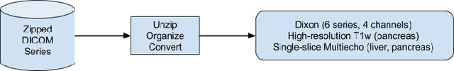
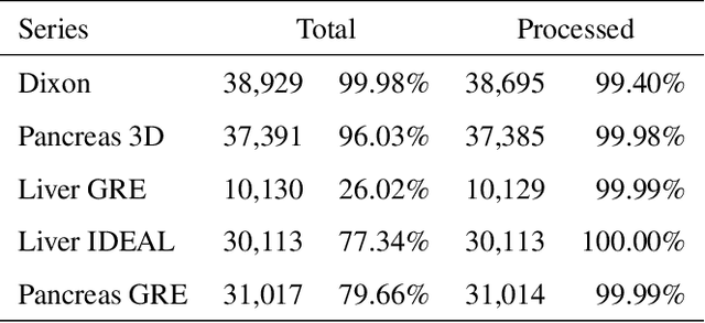
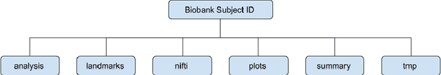
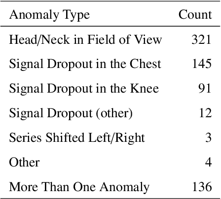
An end-to-end image analysis pipeline is presented for the abdominal MRI protocol used in the UK Biobank on the first 38,971 participants. Emphasis is on the processing steps necessary to ensure a high-level of data quality and consistency is produced in order to prepare the datasets for downstream quantitative analysis, such as segmentation and parameter estimation. Quality control procedures have been incorporated to detect and, where possible, correct issues in the raw data. Detection of fat-water swaps in the Dixon series is performed by a deep learning model and corrected automatically. Bone joints are predicted using a hybrid atlas-based registration and deep learning model for the shoulders, hips and knees. Simultaneous estimation of proton density fat fraction and transverse relaxivity (R2*) is performed using both the magnitude and phase information for the single-slice multiecho series. Approximately 98.1% of the two-point Dixon acquisitions were successfully processed and passed quality control, with 99.98% of the high-resolution T1-weighted 3D volumes succeeding. Approximately 99.98% of the single-slice multiecho acquisitions covering the liver were successfully processed and passed quality control, with 97.6% of the single-slice multiecho acquisitions covering the pancreas succeeding. At least one fat-water swap was detected in 1.8% of participants. With respect to the bone joints, approximately 3.3% of participants were missing at least one knee joint and 0.8% were missing at least one shoulder joint. For the participants who received both single-slice multiecho acquisition protocols for the liver a systematic difference between the two protocols was identified and modeled using multiple linear regression. The findings presented here will be invaluable for scientists who seek to use image-derived phenotypes from the abdominal MRI protocol.