 Add to Chrome
 Add to Firefox
 Add to Edge
Motion-guided sparse correction enables expert-quality point tracking across diverse microscopy regimes
May 28, 2026Tracking the dynamics of non-canonical biological systems in microscopy videos remains a persistent challenge. Both classical and learning-based trackers depend on expert-reviewed data to be evaluated and adapted, yet exhaustive manual annotation rarely scales to the videos where these tools are needed most. We developed RIPPLE (Refinement Interpolation Platform for Point Location Estimation), which recasts annotation as sparse correction: a user clicks a starting point, RIPPLE proposes a full trajectory, and the user intervenes only where the trajectory drifts. We tested RIPPLE on five challenging microscopy datasets from our laboratories, four from the transparent jellyfish Clytia hemisphaerica and one tracking landmarks on rapidly moving sperm. Across these, RIPPLE matched the quality of exhaustive manual annotation while reducing manual clicks by 3 to 25 times across datasets. RIPPLE thereby fills a missing layer between manual annotation and fully automated tracking, enabling immediate quantification of biological dynamics, method benchmarking, and the production of the gold-standard data needed to adapt future automated microscopy trackers.
Hypothesis-Driven Deep Learning for Out of Distribution Detection
Mar 21, 2024


Predictions of opaque black-box systems are frequently deployed in high-stakes applications such as healthcare. For such applications, it is crucial to assess how models handle samples beyond the domain of training data. While several metrics and tests exist to detect out-of-distribution (OoD) data from in-distribution (InD) data to a deep neural network (DNN), their performance varies significantly across datasets, models, and tasks, which limits their practical use. In this paper, we propose a hypothesis-driven approach to quantify whether a new sample is InD or OoD. Given a trained DNN and some input, we first feed the input through the DNN and compute an ensemble of OoD metrics, which we term latent responses. We then formulate the OoD detection problem as a hypothesis test between latent responses of different groups, and use permutation-based resampling to infer the significance of the observed latent responses under a null hypothesis. We adapt our method to detect an unseen sample of bacteria to a trained deep learning model, and show that it reveals interpretable differences between InD and OoD latent responses. Our work has implications for systematic novelty detection and informed decision-making from classifiers trained on a subset of labels.
From Hours to Seconds: Towards 100x Faster Quantitative Phase Imaging via Differentiable Microscopy
May 23, 2022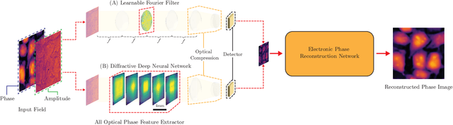
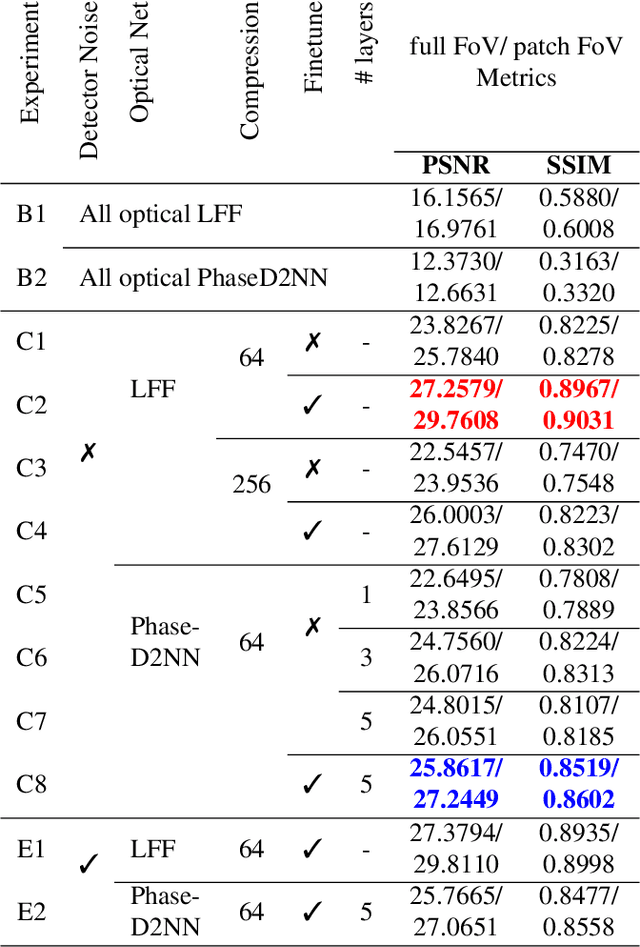
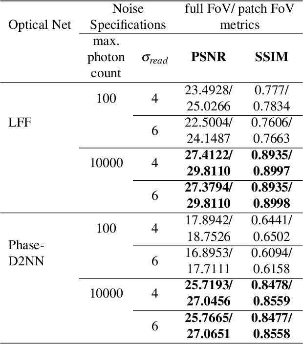
With applications ranging from metabolomics to histopathology, quantitative phase microscopy (QPM) is a powerful label-free imaging modality. Despite significant advances in fast multiplexed imaging sensors and deep-learning-based inverse solvers, the throughput of QPM is currently limited by the speed of electronic hardware. Complementarily, to improve throughput further, here we propose to acquire images in a compressed form such that more information can be transferred beyond the existing electronic hardware bottleneck. To this end, we present a learnable optical compression-decompression framework that learns content-specific features. The proposed differentiable optical-electronic quantitative phase microscopy ($\partial \mu$) first uses learnable optical feature extractors as image compressors. The intensity representation produced by these networks is then captured by the imaging sensor. Finally, a reconstruction network running on electronic hardware decompresses the QPM images. The proposed system achieves compression of $\times$ 64 while maintaining the SSIM of $\sim 0.90$ and PSNR of $\sim 30$ dB. The promising results demonstrated by our experiments open up a new pathway for achieving end-to-end optimized (i.e., optics and electronic) compact QPM systems that provide unprecedented throughput improvements.
Differentiable Microscopy Designs an All Optical Quantitative Phase Microscope
Apr 13, 2022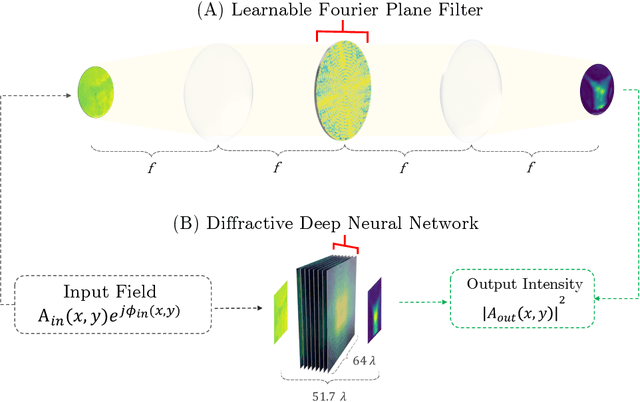
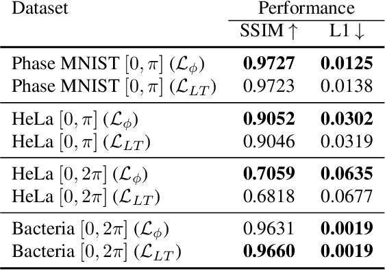

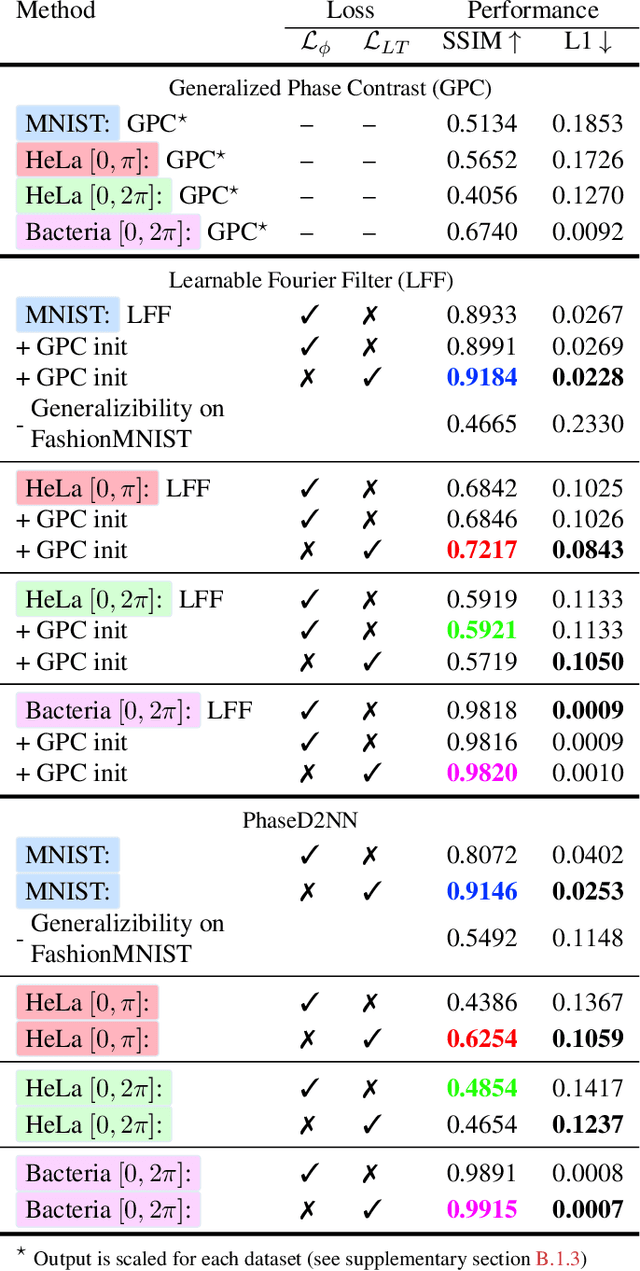
Ever since the first microscope by Zacharias Janssen in the late 16th century, scientists have been inventing new types of microscopes for various tasks. Inventing a novel architecture demands years, if not decades, worth of scientific experience and creativity. In this work, we introduce Differentiable Microscopy ($\partial\mu$), a deep learning-based design paradigm, to aid scientists design new interpretable microscope architectures. Differentiable microscopy first models a common physics-based optical system however with trainable optical elements at key locations on the optical path. Using pre-acquired data, we then train the model end-to-end for a task of interest. The learnt design proposal can then be simplified by interpreting the learnt optical elements. As a first demonstration, based on the optical 4-$f$ system, we present an all-optical quantitative phase microscope (QPM) design that requires no computational post-reconstruction. A follow-up literature survey suggested that the learnt architecture is similar to the generalized phase concept developed two decades ago. We then incorporate the generalized phase contrast concept to simplify the learning procedure. Furthermore, this physical optical setup is miniaturized using a diffractive deep neural network (D2NN). We outperform the existing benchmark for all-optical phase-to-intensity conversion on multiple datasets, and ours is the first demonstration of its kind on D2NNs. The proposed differentiable microscopy framework supplements the creative process of designing new optical systems and would perhaps lead to unconventional but better optical designs.