 Add to Chrome
 Add to Firefox
 Add to Edge
A Comprehensive Review on Digital Image Watermarking
Jul 07, 2022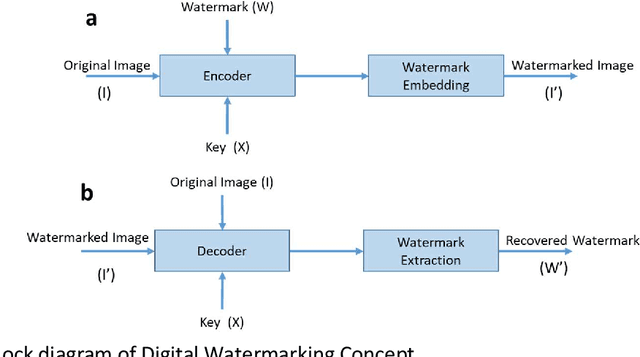
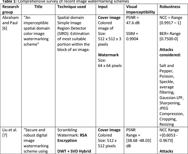
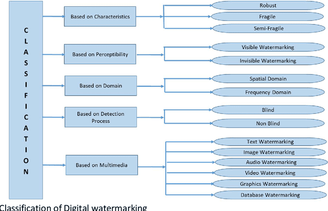
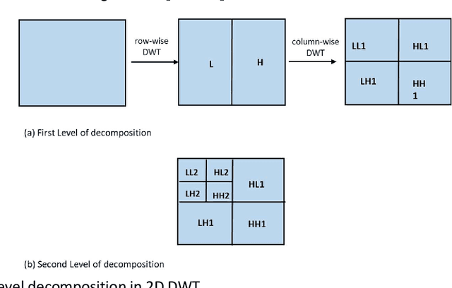
The advent of the Internet led to the easy availability of digital data like images, audio, and video. Easy access to multimedia gives rise to the issues such as content authentication, security, copyright protection, and ownership identification. Here, we discuss the concept of digital image watermarking with a focus on the technique used in image watermark embedding and extraction of the watermark. The detailed classification along with the basic characteristics, namely visual imperceptibility, robustness, capacity, security of digital watermarking is also presented in this work. Further, we have also discussed the recent application areas of digital watermarking such as healthcare, remote education, electronic voting systems, and the military. The robustness is evaluated by examining the effect of image processing attacks on the signed content and the watermark recoverability. The authors believe that the comprehensive survey presented in this paper will help the new researchers to gather knowledge in this domain. Further, the comparative analysis can enkindle ideas to improve upon the already mentioned techniques.
Biomarker Gene Identification for Breast Cancer Classification
Nov 29, 2021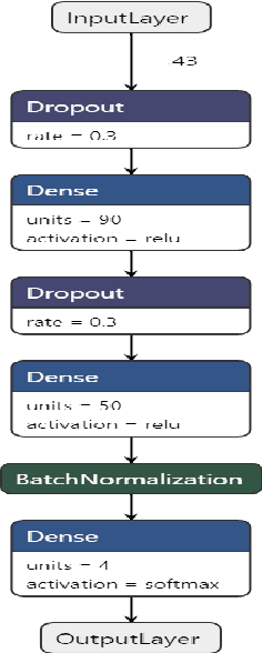
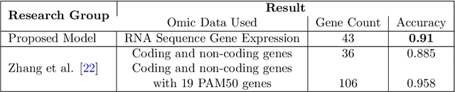
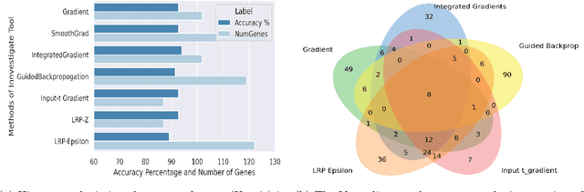
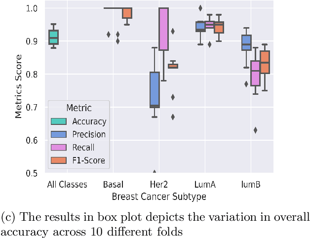
BACKGROUND: Breast cancer has emerged as one of the most prevalent cancers among women leading to a high mortality rate. Due to the heterogeneous nature of breast cancer, there is a need to identify differentially expressed genes associated with breast cancer subtypes for its timely diagnosis and treatment. OBJECTIVE: To identify a small gene set for each of the four breast cancer subtypes that could act as its signature, the paper proposes a novel algorithm for gene signature identification. METHODS: The present work uses interpretable AI methods to investigate the predictions made by the deep neural network employed for subtype classification to identify biomarkers using the TCGA breast cancer RNA Sequence data. RESULTS: The proposed algorithm led to the discovery of a set of 43 differentially expressed gene signatures. We achieved a competitive average 10-fold accuracy of 0.91, using neural network classifier. Further, gene set analysis revealed several relevant pathways, such as GRB7 events in ERBB2 and p53 signaling pathway. Using the Pearson correlation matrix, we noted that the subtype-specific genes are correlated within each subtype. CONCLUSIONS: The proposed technique enables us to find a concise and clinically relevant gene signature set.
COV-ELM classifier: An Extreme Learning Machine based identification of COVID-19 using Chest X-Ray Images
Aug 15, 2020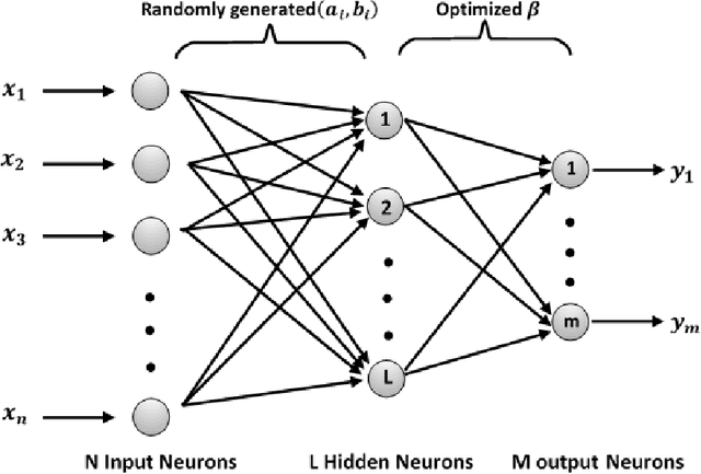
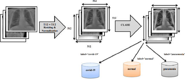
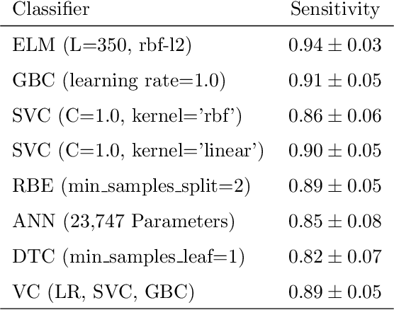
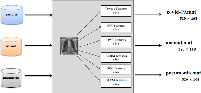
Coronaviruses constitute a family of virus that gives rise to respiratory diseases. Coronavirus disease 2019 (COVID-19) is an infectious disease caused by a newly discovered coronavirus also termed as Severe acute respiratory syndrome coronavirus 2 (SARS-CoV-2). Due to its rapid spread, WHO has declared COVID-19 outbreak a pandemic on 11th March 2020. Reverse transcription-polymerase chain reaction (RT-PCR) test is popularly used worldwide for the detection of COVID-19. However, due to the high false-negative rate of RT-PCR test, chest X-ray (CXR) imaging is emerging as a feasible alternative for the detection of COVID-19. In this work, we propose a multiclass classification model COV-ELM, based on the extreme learning machine which classifies the CXR images into one of the three classes, namely COVID-19, normal, and pneumonia. The choice of ELM in this work has been motivated by its significantly short training time as compared to conventional gradient-based learning algorithms. After some preprocessing, we extract a pool of features based on texture and frequency. This pool of features serves as an input to the ELM and a 10-fold cross-validation method is employed to evaluate the proposed model. For experimentation, we use chest X-ray (CXR) images from three publicly available sources. The results of applying COV-ELM on test data are quite promising. The COV-ELM achieved a macro average F1-score of 0.95 and the overall sensitivity of ${0.94 \pm 0.02}$ at 95% confidence interval. When compared to state-of-the-art machine learning algorithms, the COV-ELM is found to outperform in a three-class classification scenario. The main advantage of COV-ELM is that its training time being quite low, as bigger and diverse datasets become available, it can be quickly retrained as compared to its gradient-based competitor models.