 Add to Chrome
 Add to Firefox
 Add to Edge
Automatic dental superimposition of 3D intraorals and 2D photographs for human identification
Apr 07, 2026Dental comparison is considered a primary identification method, at the level of fingerprints and DNA profiling. One crucial but time-consuming step of this method is the morphological comparison. One of the main challenges to apply this method is the lack of ante-mortem medical records, specially on scenarios such as migrant death at the border and/or in countries where there is no universal healthcare. The availability of photos on social media where teeth are visible has led many odontologists to consider morphological comparison using them. However, state-of-the-art proposals have significant limitations, including the lack of proper modeling of perspective distortion and the absence of objective approaches that quantify morphological differences. Our proposal involves a 3D (post-mortem scan) - 2D (ante-mortem photos) approach. Using computer vision and optimization techniques, we replicate the ante-mortem image with the 3D model to perform the morphological comparison. Two automatic approaches have been developed: i) using paired landmarks and ii) using a segmentation of the teeth region to estimate camera parameters. Both are capable of obtaining very promising results over 20,164 cross comparisons from 142 samples, obtaining mean ranking values of 1.6 and 1.5, respectively. These results clearly outperform filtering capabilities of automatic dental chart comparison approaches, while providing an automatic, objective and quantitative score of the morphological correspondence, easily to interpret and analyze by visualizing superimposed images.
Study on the identification limits of craniofacial superimposition
Jan 24, 2023Craniofacial Superimposition involves the superimposition of an image of a skull with a number of ante-mortem face images of an individual and the analysis of their morphological correspondence. Despite being used for one century, it is not yet a mature and fully accepted technique due to the absence of solid scientific approaches, significant reliability studies, and international standards. In this paper we present a comprehensive experimentation on the limitations of Craniofacial Superimposition as a forensic identification technique. The study involves different experiments over more than 1 Million comparisons performed by a landmark-based automatic 3D/2D superimposition method. The total sample analyzed consists of 320 subjects and 29 craniofacial landmarks.
Automatic cephalometric landmarks detection on frontal faces: an approach based on supervised learning techniques
Apr 24, 2019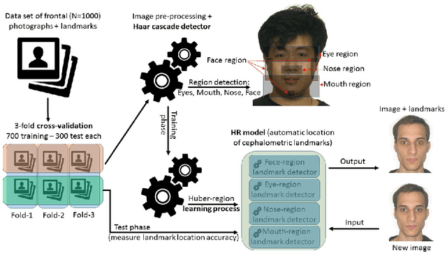
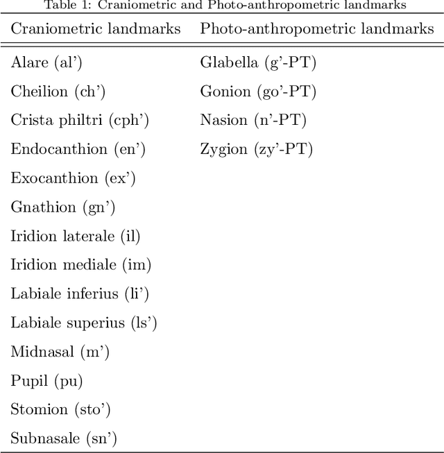
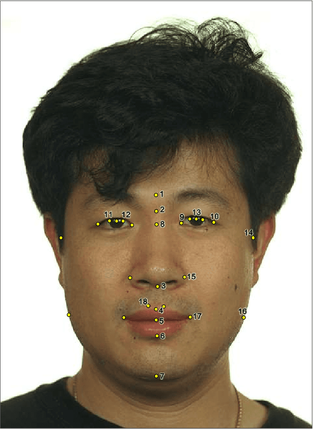
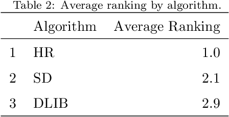
Facial landmarks are employed in many research areas such as facial recognition, craniofacial identification, age and sex estimation among the most important. In the forensic field, the focus is on the analysis of a particular set of facial landmarks, defined as cephalometric landmarks. Previous works demonstrated that the descriptive adequacy of these anatomical references for an indirect application (photo-anthropometric description) increased the marking precision of these points, contributing to a greater reliability of these analyzes. However, most of them are performed manually and all of them are subjectivity inherent to the expert examiners. In this sense, the purpose of this work is the development and validation of automatic techniques to detect cephalometric landmarks from digital images of frontal faces in forensic field. The presented approach uses a combination of computer vision and image processing techniques within a supervised learning procedures. The proposed methodology obtains similar precision to a group of human manual cephalometric reference markers and result to be more accurate against others state-of-the-art facial landmark detection frameworks. It achieves a normalized mean distance (in pixel) error of 0.014, similar to the mean inter-expert dispersion (0.009) and clearly better than other automatic approaches also analyzed along of this work (0.026 and 0.101).