 Add to Chrome
 Add to Firefox
 Add to Edge
Attention-Enhanced Deep Learning Ensemble for Breast Density Classification in Mammography
Jul 08, 2025Breast density assessment is a crucial component of mammographic interpretation, with high breast density (BI-RADS categories C and D) representing both a significant risk factor for developing breast cancer and a technical challenge for tumor detection. This study proposes an automated deep learning system for robust binary classification of breast density (low: A/B vs. high: C/D) using the VinDr-Mammo dataset. We implemented and compared four advanced convolutional neural networks: ResNet18, ResNet50, EfficientNet-B0, and DenseNet121, each enhanced with channel attention mechanisms. To address the inherent class imbalance, we developed a novel Combined Focal Label Smoothing Loss function that integrates focal loss, label smoothing, and class-balanced weighting. Our preprocessing pipeline incorporated advanced techniques, including contrast-limited adaptive histogram equalization (CLAHE) and comprehensive data augmentation. The individual models were combined through an optimized ensemble voting approach, achieving superior performance (AUC: 0.963, F1-score: 0.952) compared to any single model. This system demonstrates significant potential to standardize density assessments in clinical practice, potentially improving screening efficiency and early cancer detection rates while reducing inter-observer variability among radiologists.
Joint brain tumor segmentation from multi MR sequences through a deep convolutional neural network
Mar 07, 2022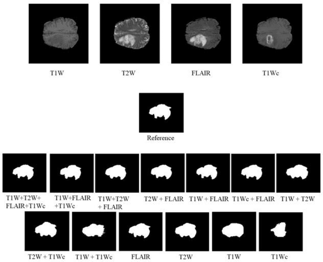
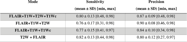
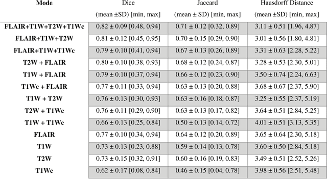
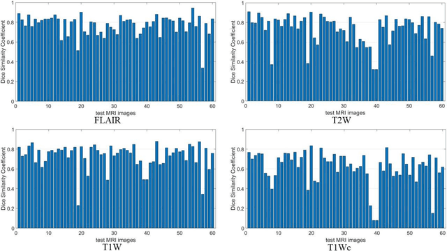
Brain tumor segmentation is highly contributive in diagnosing and treatment planning. The manual brain tumor delineation is a time-consuming and tedious task and varies depending on the radiologists skill. Automated brain tumor segmentation is of high importance, and does not depend on either inter or intra-observation. The objective of this study is to automate the delineation of brain tumors from the FLAIR, T1 weighted, T2 weighted, and T1 weighted contrast-enhanced MR sequences through a deep learning approach, with a focus on determining which MR sequence alone or which combination thereof would lead to the highest accuracy therein.
Quantitative analysis of image quality in low-dose CT imaging for Covid-19 patients
Feb 16, 2021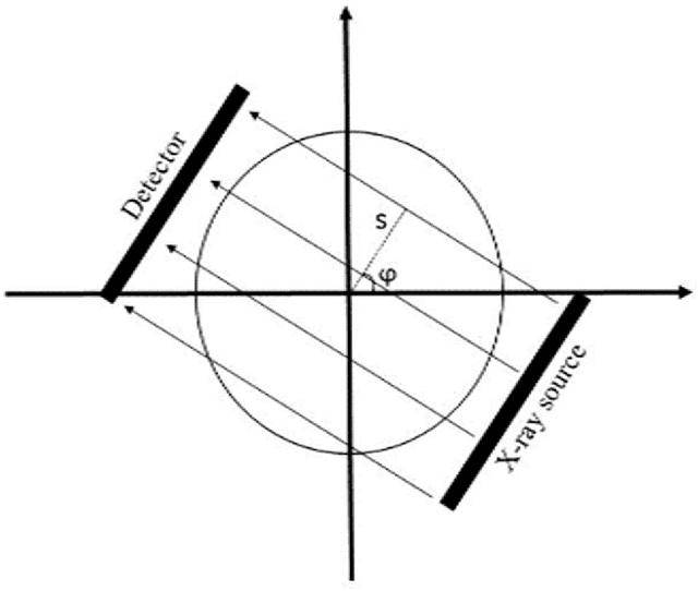
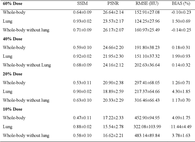
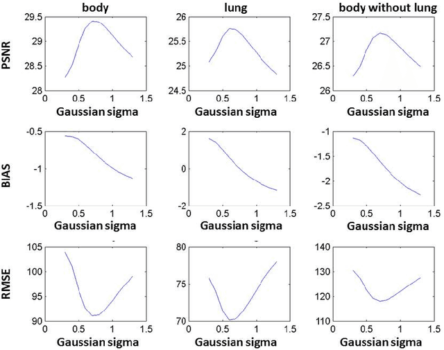
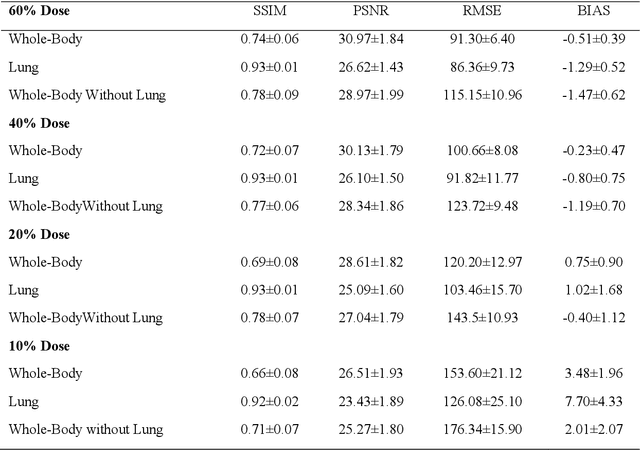
We set out to simulate four reduced dose-levels (60%-dose, 40%-dose, 20%-dose, and 10%-dose) of standard CT imaging using Beer-Lambert's law across 49 patients infected with COVID-19. Then, three denoising filters, namely Gaussian, Bilateral, and Median, were applied to the different low-dose CT images, the quality of which was assessed prior to and after the application of the various filters via calculation of peak signal-to-noise ratio (PSNR), root mean square error (RMSE), structural similarity index measure (SSIM), and relative CT-value bias, separately for the lung tissue and whole-body. The quantitative evaluation indicated that 10%-dose CT images have inferior quality (with RMSE=322.1-+104.0 HU and bias=11.44-+4.49% in the lung) even after the application of the denoising filters. The bilateral filter exhibited superior performance to suppress the noise and recover the underlying signals in low-dose CT images compared to the other denoising techniques. The bilateral filter led to RMSE and bias of 100.21-+16.47 HU, -0.21-+1.20%, respectively in the lung regions for 20%-dose CT images compared to the Gaussian filter with RMSE=103.46-+15.70 HU and bias=1.02-+1.68%, median filter with RMSE=129.60-+18.09 HU and bias=-6.15-+2.24%, and the nonfiltered 20%-dose CT with RMSE=217.37-+64.66 HU and bias=4.30-+1.85%. In conclusion, the 20%-dose CT imaging followed by the bilateral filtering introduced a reasonable compromise between image quality and patient dose reduction.