 Add to Chrome
 Add to Firefox
 Add to Edge
Generative Deep Learning for Computational Destaining and Restaining of Unregistered Digital Pathology Images
May 14, 2026Conditional generative adversarial networks (cGANs) have enabled high-fidelity computational staining and destaining of hematoxylin and eosin (H&E) in digital pathology whole-slide images (WSI). However, their ability to generalize to out-of-distribution WSI across institutions without retraining remains insufficiently characterized. Previously developed cGAN models trained on 102 registered prostate core biopsy WSIs from Brigham and Women's Hospital were evaluated on 82 spatially unregistered WSIs acquired at Stanford University. To mitigate domain shift without retraining, a preprocessing pipeline consisting of histogram-based stain normalization for H&E-stained WSIs and channel-wise intensity calibration for unstained WSIs was developed. Because image registration was intentionally omitted for real-world deployment conditions, the reported quantitative results are conservative lower bounds reflecting both model performance and limited spatial alignment. Under these conditions, virtual destaining achieved a Pearson correlation coefficient (PCC) of 0.854, structural similarity index measure (SSIM) of 0.699, and peak signal-to-noise ratio (PSNR) of 18.41 dB. H&E restaining from computationally destained outputs outperformed direct staining from ground-truth unstained inputs across all metrics (PCC: 0.798 vs. 0.715; SSIM: 0.756 vs. 0.718; PSNR: 20.08 vs. 18.51 dB), suggesting that preprocessing quality may be more limiting than model capacity. Qualitative pathological review indicated preservation of benign glandular structures while showing that malignant glands were often rendered with vessel-like morphologies. These findings support the feasibility of applying cGAN-based computational H&E staining and destaining generative models to external WSI datasets using preprocessing-based adaptation alone while defining specific morphological targets for future domain adaptation.
High Accuracy Tumor Diagnoses and Benchmarking of Hematoxylin and Eosin Stained Prostate Core Biopsy Images Generated by Explainable Deep Neural Networks
Aug 02, 2019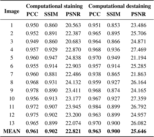
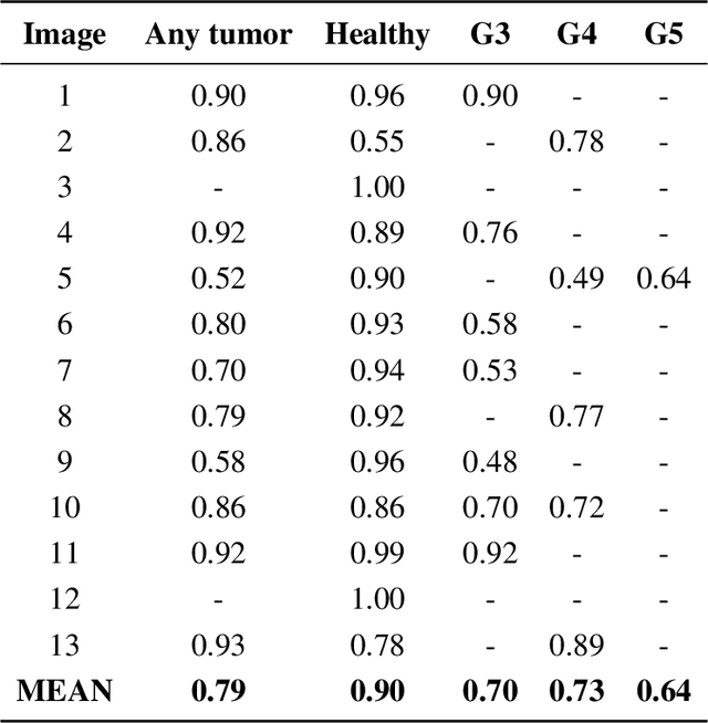
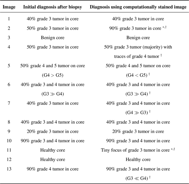
Histopathological diagnoses of tumors in tissue biopsy after Hematoxylin and Eosin (H&E) staining is the gold standard for oncology care. H&E staining is slow and uses dyes, reagents and precious tissue samples that cannot be reused. Thousands of native nonstained RGB Whole Slide Image (RWSI) patches of prostate core tissue biopsies were registered with their H&E stained versions. Conditional Generative Adversarial Neural Networks (cGANs) that automate conversion of native nonstained RWSI to computational H&E stained images were then trained. High similarities between computational and H&E dye stained images with Structural Similarity Index (SSIM) 0.902, Pearsons Correlation Coefficient (CC) 0.962 and Peak Signal to Noise Ratio (PSNR) 22.821 dB were calculated. A second cGAN performed accurate computational destaining of H&E dye stained images back to their native nonstained form with SSIM 0.9, CC 0.963 and PSNR 25.646 dB. A single-blind study computed more than 95% pixel-by-pixel overlap between prostate tumor annotations on computationally stained images, provided by five-board certified MD pathologists, with those on H&E dye stained counterparts. We report the first visualization and explanation of neural network kernel activation maps during H&E staining and destaining of RGB images by cGANs. High similarities between kernel activation maps of computational and H&E stained images (Mean-Squared Errors <0.0005) provide additional mathematical and mechanistic validation of the staining system. Our neural network framework thus is automated, explainable and performs high precision H&E staining and destaining of low cost native RGB images, and is computer vision and physician authenticated for rapid and accurate tumor diagnoses.