 Add to Chrome
 Add to Firefox
 Add to Edge
Retinal Vessel Segmentation Using the 2-D Morlet Wavelet and Supervised Classification
Paper and Code
May 11, 2006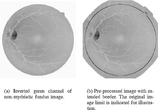
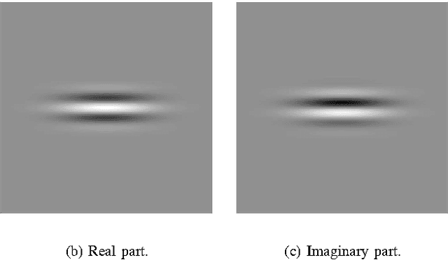
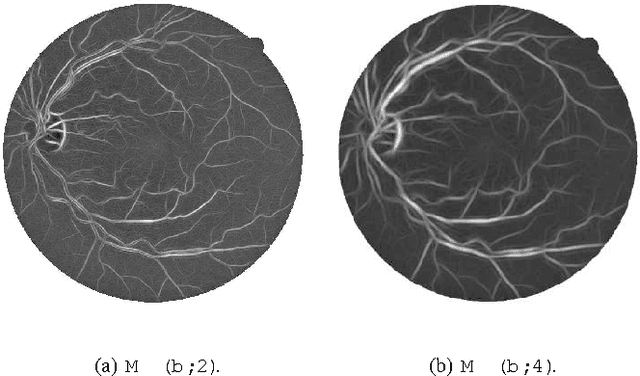
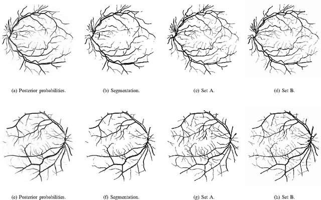
We present a method for automated segmentation of the vasculature in retinal images. The method produces segmentations by classifying each image pixel as vessel or non-vessel, based on the pixel's feature vector. Feature vectors are composed of the pixel's intensity and continuous two-dimensional Morlet wavelet transform responses taken at multiple scales. The Morlet wavelet is capable of tuning to specific frequencies, thus allowing noise filtering and vessel enhancement in a single step. We use a Bayesian classifier with class-conditional probability density functions (likelihoods) described as Gaussian mixtures, yielding a fast classification, while being able to model complex decision surfaces and compare its performance with the linear minimum squared error classifier. The probability distributions are estimated based on a training set of labeled pixels obtained from manual segmentations. The method's performance is evaluated on publicly available DRIVE and STARE databases of manually labeled non-mydriatic images. On the DRIVE database, it achieves an area under the receiver operating characteristic (ROC) curve of 0.9598, being slightly superior than that presented by the method of Staal et al.