 Add to Chrome
 Add to Firefox
 Add to Edge
Ultrafast Ultrasound Imaging for 3D Shear Wave Absolute Vibro-Elastography
Paper and Code
Mar 26, 2022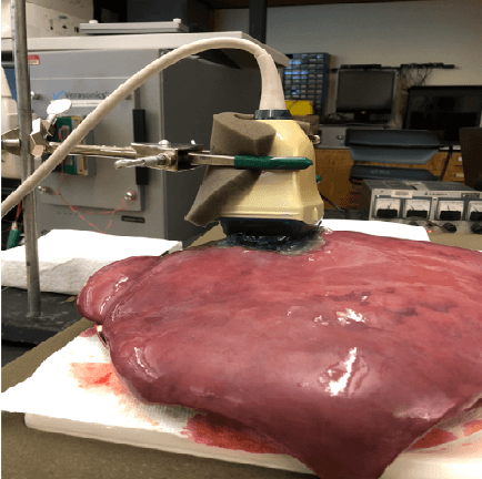
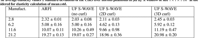
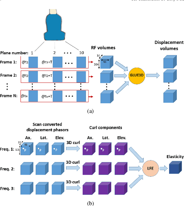
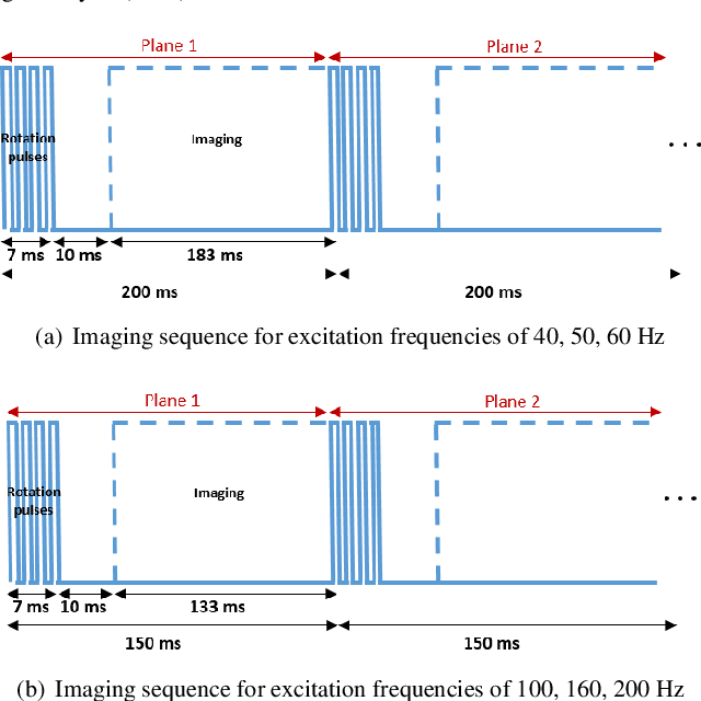
Shear wave absolute vibro-elastography (S-WAVE) is an imaging technique that generates steady-state shear waves inside the tissue using multi-frequency excitation from an external vibration source. In this work, plane wave imaging is introduced to reduce total acquisition time while retaining the benefit of 3D formulation. Plane wave imaging with a frame rate of 3000 frames/s is followed by 3D absolute elasticity estimation. We design two imaging sequences of ultrafast S-WAVE for two sets of excitation frequencies using a Verasonics system and a motorized swept ultrasound transducer to synchronize ultrasound acquisition with the external mechanical excitation. The overall data collection time is improved by 83-88% compared to the original 3D S-WAVE because of the per-channel acquisition offered by the Verasonics system. Tests are performed on liver fibrosis tissue-mimicking phantoms and on ex vivo bovine liver. The curl operator was previously used in magnetic resonance elastography (MRE) to cancel out the effect of the compressional waves. In this work, we apply the curl operator to the full 3D displacement field followed by 3D elasticity reconstruction. The results of phantom experiment show more accurate elasticity estimation as well as 18% less standard deviation (STD) compared to reconstruction using the curl of a 2D displacement field and 45% less STD than without the curl. We also compare our experimental results with a previous method based on acoustic radiation force impulse (ARFI) and achieve closer results to phantom manufacturer provided values with ultrafast S-WAVE. Furthermore, the dependency of the bovine liver elasticity on the frequency of excitation was also shown with our system.