Get our free extension to see links to code for papers anywhere online!Free add-on: code for papers everywhere!Free add-on: See code for papers anywhere!
 Add to Chrome
 Add to Firefox
 Add to Edge
Automated image segmentation for detecting cell spreading for metastasizing assessments of cancer development
Paper and Code
Jan 01, 2018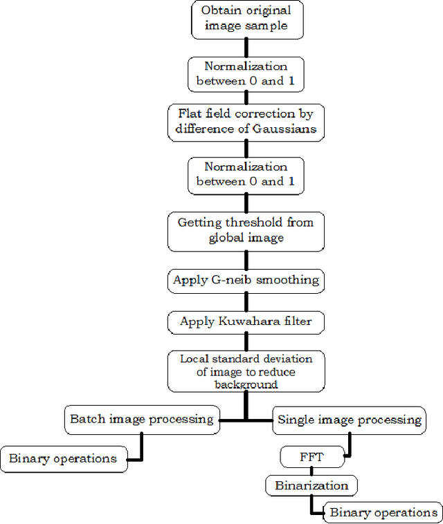
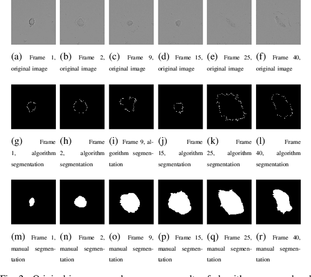
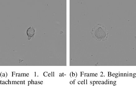
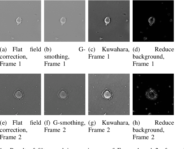
The automated segmentation of cells in microscopic images is an open research problem that has important implications for studies of the developmental and cancer processes based on in vitro models. In this paper, we present the approach for segmentation of the DIC images of cultured cells using G-neighbor smoothing followed by Kauwahara filtering and local standard deviation approach for boundary detection. NIH FIJI/ImageJ tools are used to create the ground truth dataset. The results of this work indicate that detection of cell boundaries using segmentation approach even in the case of realistic measurement conditions is a challenging problem.
* 2017 International Conference on Advances in Computing,
Communications and Informatics (ICACCI), Udupi, 2017, pp. 2382-2387
View paper on