 Add to Chrome
 Add to Firefox
 Add to Edge
EA-LiteUNet: An Edge-Adaptive and Resource-Efficient U-Net for Boundary-Sensitive Dermoscopic Image Segmentation
Aug 16, 2026
Accurate boundary delineation remains a persistent challenge in dermoscopic image segmentation because of blurred lesion margins, heterogeneous textures, and complex background artifacts. From a signal-processing perspective, lesion boundaries represent high-frequency components that are highly susceptible to aliasing, noise amplification, and information loss. Consequently, repeated downsampling and feature transformations in conventional convolutional architectures often lead to severely degraded boundary representations. To address these limitations, we propose EA-LiteUNet, an edge-adaptive and computationally efficient U-Net variant specifically designed for boundary-sensitive medical image segmentation. The architecture integrates three core mechanisms: (1) boundary-aware representation learning to suppress aliasing and preserve high-frequency structural details; (2) attention-guided feature modulation to selectively enhance boundary-relevant responses across multi-scale features; and (3) a resource-adaptive inference strategy to dynamically balance segmentation accuracy and computational efficiency. Extensive evaluations across three public dermoscopic datasets demonstrate that EA-LiteUNet consistently achieves superior boundary precision. Specifically, on the ISIC 2018 dataset, the method significantly reduces the 95% Hausdorff Distance (HD95) to 12.89 pixels while maintaining a robust Dice score of 92.08%. Notably, this strong performance is achieved with an ultralightweight configuration of merely 0.29M parameters and 1.17 GFLOPs. Ablation studies further validate the complementary effects of these components, confirming their contribution to enhanced boundary fidelity and stable optimization.

A Comprehensive Review of U-Net and Its Variants: Advances and Applications in Medical Image Segmentation
Feb 09, 2025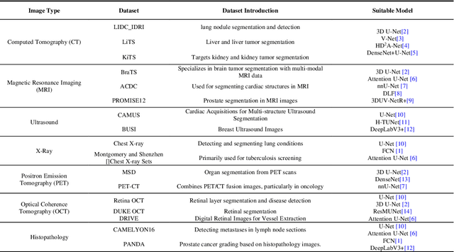
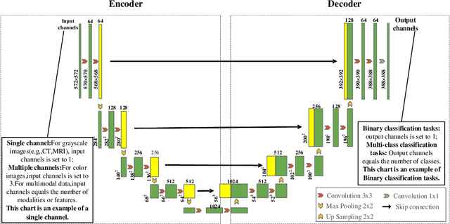
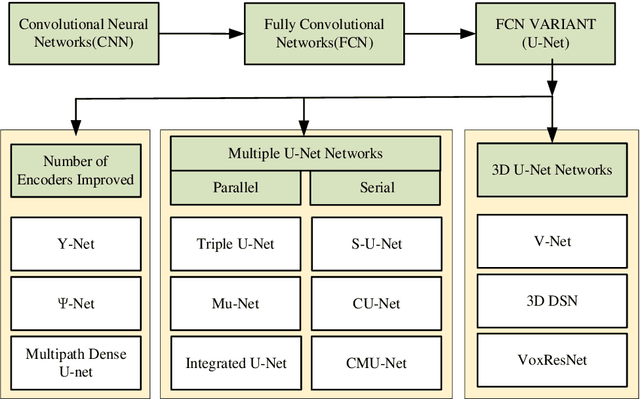
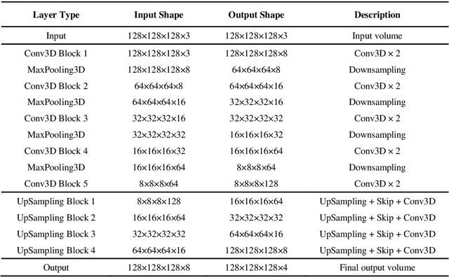
Medical images often exhibit low and blurred contrast between lesions and surrounding tissues, with considerable variation in lesion edges and shapes even within the same disease, leading to significant challenges in segmentation. Therefore, precise segmentation of lesions has become an essential prerequisite for patient condition assessment and formulation of treatment plans. Significant achievements have been made in research related to the U-Net model in recent years. It improves segmentation performance and is extensively applied in the semantic segmentation of medical images to offer technical support for consistent quantitative lesion analysis methods. First, this paper classifies medical image datasets on the basis of their imaging modalities and then examines U-Net and its various improvement models from the perspective of structural modifications. The research objectives, innovative designs, and limitations of each approach are discussed in detail. Second, we summarize the four central improvement mechanisms of the U-Net and U-Net variant algorithms: the jump-connection mechanism, residual-connection mechanism, 3D-UNet, and transformer mechanism. Finally, we examine the relationships among the four core enhancement mechanisms and commonly utilized medical datasets and propose potential avenues and strategies for future advancements. This paper provides a systematic summary and reference for researchers in related fields, and we look forward to designing more efficient and stable medical image segmentation network models based on the U-Net network.