 Add to Chrome
 Add to Firefox
 Add to Edge
Towards order of magnitude X-ray dose reduction in breast cancer imaging using phase contrast and deep denoising
May 09, 2025


Breast cancer is the most frequently diagnosed human cancer in the United States at present. Early detection is crucial for its successful treatment. X-ray mammography and digital breast tomosynthesis are currently the main methods for breast cancer screening. However, both have known limitations in terms of their sensitivity and specificity to breast cancers, while also frequently causing patient discomfort due to the requirement for breast compression. Breast computed tomography is a promising alternative, however, to obtain high-quality images, the X-ray dose needs to be sufficiently high. As the breast is highly radiosensitive, dose reduction is particularly important. Phase-contrast computed tomography (PCT) has been shown to produce higher-quality images at lower doses and has no need for breast compression. It is demonstrated in the present study that, when imaging full fresh mastectomy samples with PCT, deep learning-based image denoising can further reduce the radiation dose by a factor of 16 or more, without any loss of image quality. The image quality has been assessed both in terms of objective metrics, such as spatial resolution and contrast-to-noise ratio, as well as in an observer study by experienced medical imaging specialists and radiologists. This work was carried out in preparation for live patient PCT breast cancer imaging, initially at specialized synchrotron facilities.
Single-exposure elemental differentiation and texture-sensitive phase-retrieval imaging with a neutron counting micro-channel plate detector
Nov 09, 2023


Micro-channel plate (MCP) detectors, when used at pulsed-neutron-source instruments, offer the possibility of high spatial resolution and high contrast imaging with pixel-level spectroscopic information. Here we demonstrate the possibility of multimodal analysis including total neutron cross-section spectra measurements, quantitative material differentiation imaging and texture-sensitive in-line phase imaging, from a single exposure using an MCP detector. This multimodal approach operates in full-field imaging mode, with the neutron transmission spectra acquired at each individual detector pixel. Due to the polychromatic nature of the beam and spectroscopic resolving capability of the detector, no energy scanning is required. Good agreement with the library reference data is demonstrated for neutron cross-section spectra measurements. Two different images corresponding to two selected energy bandwidths are used for elemental differentiation imaging. Moreover, the presence of changes in texture, i.e., preferred grain orientation, in the sample is identified from our phase-retrieval imaging results.
Tomographic phase and attenuation extraction for a sample composed of unknown materials using X-ray propagation-based phase-contrast imaging
Oct 14, 2021
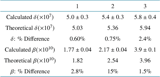
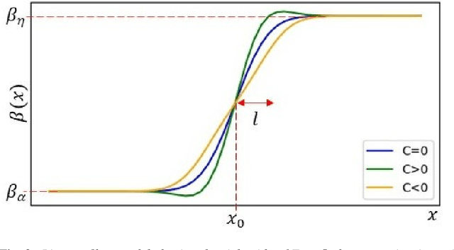
Propagation-based phase-contrast X-ray imaging (PB-PCXI) generates image contrast by utilizing sample-imposed phase-shifts. This has proven useful when imaging weakly-attenuating samples, as conventional attenuation-based imaging does not always provide adequate contrast. We present a PB-PCXI algorithm capable of extracting the X-ray attenuation, $\beta$, and refraction, $\delta$, components of the complex refractive index of distinct materials within an unknown sample. The method involves curve-fitting an error-function-based model to a phase-retrieved interface in a PB-PCXI tomographic reconstruction, which is obtained when Paganin-type phase-retrieval is applied with incorrect values of $\delta$ and $\beta$. The fit parameters can then be used to calculate true $\delta$ and $\beta$ values for composite materials. This approach requires no a priori sample information, making it broadly applicable. Our PB-PCXI reconstruction is single distance, requiring only one exposure per tomographic angle, which is important for radiosensitive samples. We apply this approach to a breast-tissue sample, recovering the refraction component, $\delta$, with 0.6 - 2.4\% accuracy compared to theoretical values.