 Add to Chrome
 Add to Firefox
 Add to Edge
IdentiARAT: Toward Automated Identification of Individual ARAT Items from Wearable Sensors
Apr 17, 2025

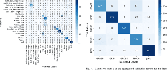

This study explores the potential of using wrist-worn inertial sensors to automate the labeling of ARAT (Action Research Arm Test) items. While the ARAT is commonly used to assess upper limb motor function, its limitations include subjectivity and time consumption of clinical staff. By using IMU (Inertial Measurement Unit) sensors and MiniROCKET as a time series classification technique, this investigation aims to classify ARAT items based on sensor recordings. We test common preprocessing strategies to efficiently leverage included information in the data. Afterward, we use the best preprocessing to improve the classification. The dataset includes recordings of 45 participants performing various ARAT items. Results show that MiniROCKET offers a fast and reliable approach for classifying ARAT domains, although challenges remain in distinguishing between individual resembling items. Future work may involve improving classification through more advanced machine-learning models and data enhancements.
MRI lung lobe segmentation in pediatric cystic fibrosis patients using a recurrent neural network trained with publicly accessible CT datasets
Aug 31, 2021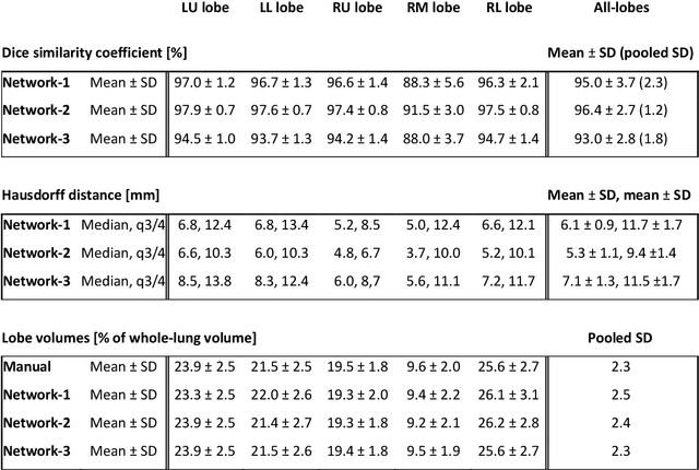
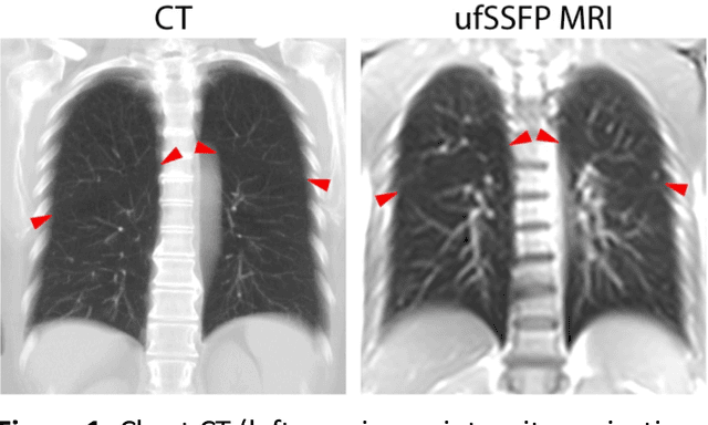
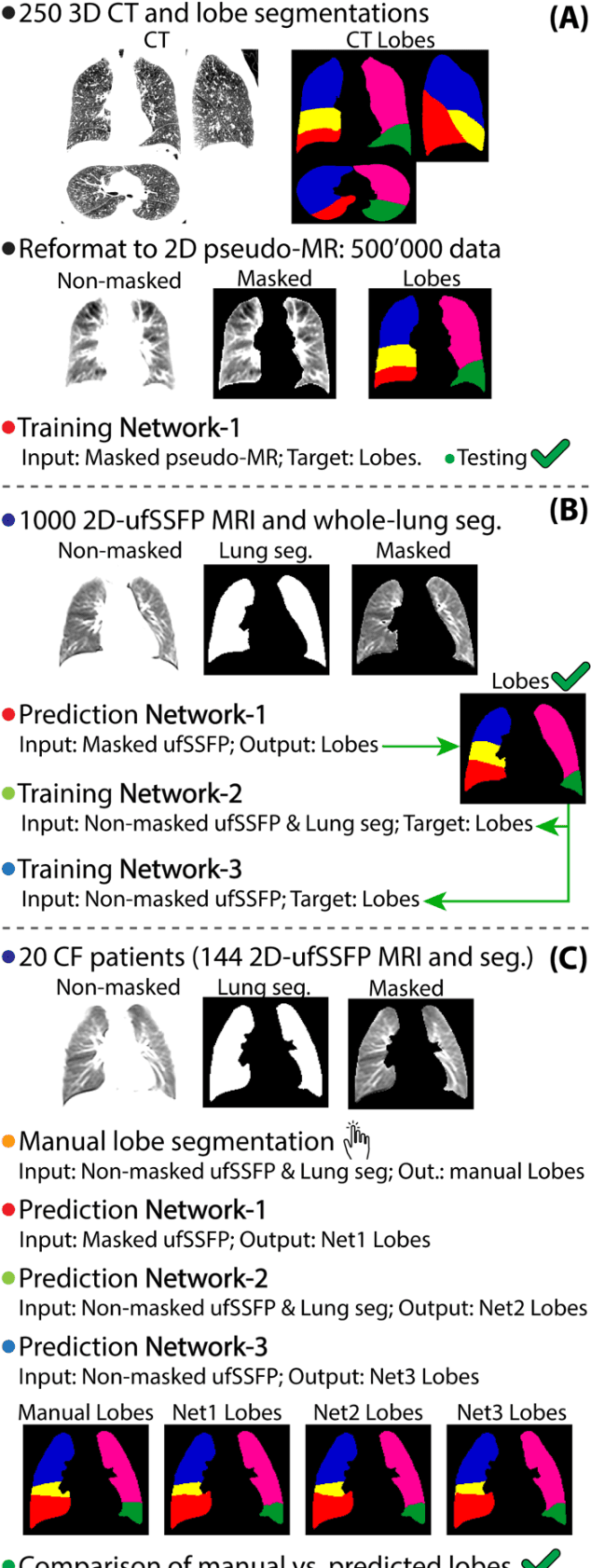
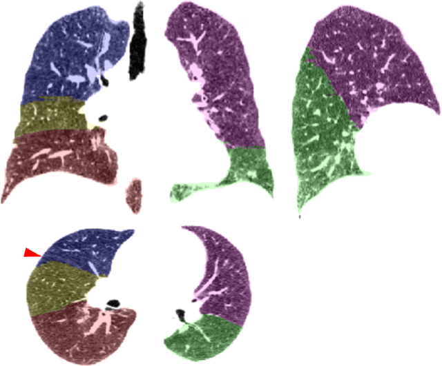
Purpose: To introduce a widely applicable workflow for pulmonary lobe segmentation of MR images using a recurrent neural network (RNN) trained with chest computed tomography (CT) datasets. The feasibility is demonstrated for 2D coronal ultra-fast balanced steady-state free precession (ufSSFP) MRI. Methods: Lung lobes of 250 publicly accessible CT datasets of adults were segmented with an open-source CT-specific algorithm. To match 2D ufSSFP MRI data of pediatric patients, both CT data and segmentations were translated into pseudo-MR images, masked to suppress anatomy outside the lung. Network-1 was trained with pseudo-MR images and lobe segmentations, and applied to 1000 masked ufSSFP images to predict lobe segmentations. These outputs were directly used as targets to train Network-2 and Network-3 with non-masked ufSSFP data as inputs, and an additional whole-lung mask as input for Network-2. Network predictions were compared to reference manual lobe segmentations of ufSSFP data in twenty pediatric cystic fibrosis patients. Manual lobe segmentations were performed by splitting available whole-lung segmentations into lobes. Results: Network-1 was able to segment the lobes of ufSSFP images, and Network-2 and Network-3 further increased segmentation accuracy and robustness. The average all-lobe Dice similarity coefficients were 95.0$\pm$2.3 (mean$\pm$pooled SD [%]), 96.4$\pm$1.2, 93.0$\pm$1.8, and the average median Hausdorff distances were 6.1$\pm$0.9 (mean$\pm$SD [mm]), 5.3$\pm$1.1, 7.1$\pm$1.3, for Network-1, Network-2, and Network-3, respectively. Conclusions: RNN lung lobe segmentation of 2D ufSSFP imaging is feasible, in good agreement with manual segmentations. The proposed workflow might provide rapid access to automated lobe segmentations for various lung MRI examinations and quantitative analyses.