 Add to Chrome
 Add to Firefox
 Add to Edge
VRXU-net: A Deep Learning Approach for Brain Ischemic Stroke Lesion Detection and Segmentation in T1W MRI
May 20, 2026
When the blood supply to the brain is obstructed by a clot, oxygen delivery to brain tissues becomes insufficient, leading to cellular necrosis. In healthcare settings, accurately identifying and delineating ischemic lesion boundaries is essential for treatment and surgical planning. However, ischemic stroke lesions vary widely in shape, size, and location, and in grayscale MRI modalities such as T1W they may resemble surrounding brain structures. This makes lesion detection and segmentation a challenging task for clinicians. This study introduces a novel VRU-Net architecture, derived from visual features, residual connections, and a U-shaped network, for detecting and segmenting ischemic stroke lesions in 3D magnetic resonance imaging scans. The proposed method first uses a modified VGG model to identify ischemic stroke in separate 2D slices. Then, a U-shaped segmentation model with residual blocks segments the lesion in each slice. This procedure is applied independently to the axial, sagittal, and coronal planes, and the final output is generated by aggregating the three segmentation results. To improve both performance and processing speed, a high-performance classifier is applied before the segmentation model in a sequential framework. This strategy reduces unnecessary segmentation of non-lesion slices and improves overall accuracy. In addition, decomposing 3D images into 2D slices reduces model complexity while allowing information from three anatomical planes to support more accurate lesion localization. The proposed model is trained on the Anatomical Tracings of Lesions After Stroke dataset and outperforms state-of-the-art models in terms of accuracy and Dice coefficient. Moreover, the segmentation output provides feedback that helps the classification model reduce false-positive predictions.

Empowering Medical Imaging with Artificial Intelligence: A Review of Machine Learning Approaches for the Detection, and Segmentation of COVID-19 Using Radiographic and Tomographic Images
Jan 13, 2024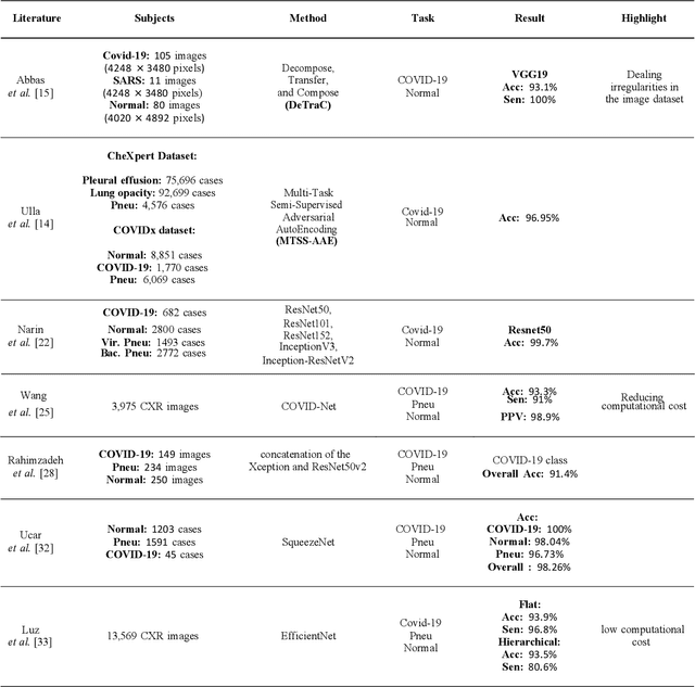
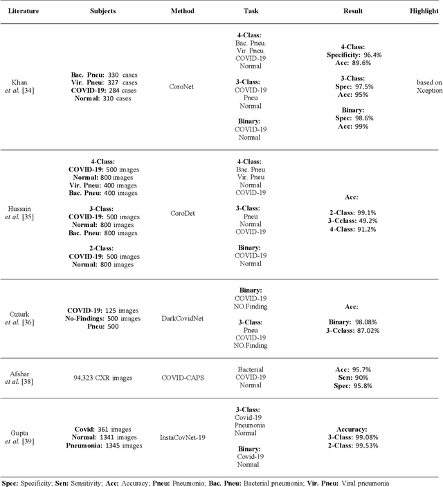
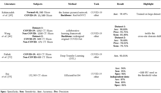
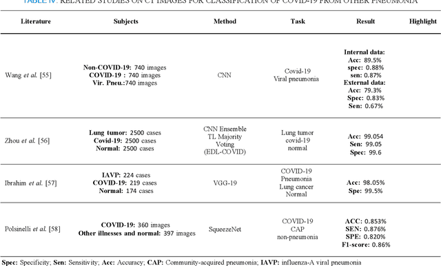
Since 2019, the global dissemination of the Coronavirus and its novel strains has resulted in a surge of new infections. The use of X-ray and computed tomography (CT) imaging techniques is critical in diagnosing and managing COVID-19. Incorporating artificial intelligence (AI) into the field of medical imaging is a powerful combination that can provide valuable support to healthcare professionals.This paper focuses on the methodological approach of using machine learning (ML) to enhance medical imaging for COVID-19 diagnosis.For example, deep learning can accurately distinguish lesions from other parts of the lung without human intervention in a matter of minutes.Moreover, ML can enhance performance efficiency by assisting radiologists in making more precise clinical decisions, such as detecting and distinguishing Covid-19 from different respiratory infections and segmenting infections in CT and X-ray images, even when the lesions have varying sizes and shapes.This article critically assesses machine learning methodologies utilized for the segmentation, classification, and detection of Covid-19 within CT and X-ray images, which are commonly employed tools in clinical and hospital settings to represent the lung in various aspects and extensive detail.There is a widespread expectation that this technology will continue to hold a central position within the healthcare sector, driving further progress in the management of the pandemic.