 Add to Chrome
 Add to Firefox
 Add to Edge
Machine learning enables experimental access to photon-by-photon arrival times in scintillation detectors
May 27, 2026
Scintillation detectors with excellent timing resolution enable more precise localization of radiation sources in positron emission tomography, leading to substantial improvements in diagnostic capability for diseases such as cancer and dementia. At the extreme timing precision required for such applications at the picosecond scale, detector performance is governed by the microscopic dynamics of scintillation photons generated within the detector and their subsequent detection processes. However, detector signals have conventionally been treated only as collective responses of many photons due to structural constraints inherent to photodetectors. In this study, we overcome this fundamental limitation using deep learning, enabling direct access to the timing information of individual photons. The proposed method estimates photon-by-photon arrival times directly from detector waveforms without requiring any modification to the detector structure; the method operates on an event-by-event basis without ground-truth labels by integrating an unsupervised learning framework with a physically informed detector-response model. Through comprehensive validation combining Monte Carlo simulation and experimental measurements across various detector configurations, we experimentally demonstrate improved timing resolution, visualized depth-of-interaction-dependent photon transport, and classified Cherenkov and scintillation photons based on the estimated photon-level timing information using a unified deep learning-based framework. These results provide experimental access to photon dynamics, bridging the gap between theoretical modeling and experimental observation, and they open a new data-driven pathway for discovery in detector physics and optimization.

Anatomical-Guided Attention Enhances Unsupervised PET Image Denoising Performance
Sep 08, 2021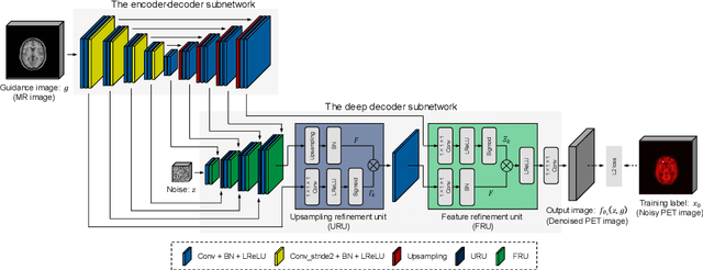

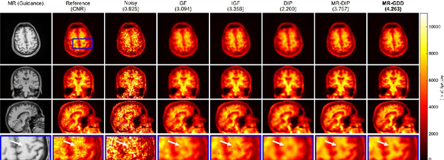
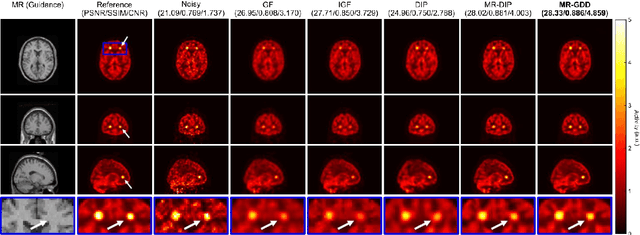
Although supervised convolutional neural networks (CNNs) often outperform conventional alternatives for denoising positron emission tomography (PET) images, they require many low- and high-quality reference PET image pairs. Herein, we propose an unsupervised 3D PET image denoising method based on an anatomical information-guided attention mechanism. The proposed magnetic resonance-guided deep decoder (MR-GDD) utilizes the spatial details and semantic features of MR-guidance image more effectively by introducing encoder-decoder and deep decoder subnetworks. Moreover, the specific shapes and patterns of the guidance image do not affect the denoised PET image, because the guidance image is input to the network through an attention gate. In a Monte Carlo simulation of [$^{18}$F]fluoro-2-deoxy-D-glucose (FDG), the proposed method achieved the highest peak signal-to-noise ratio and structural similarity (27.92 $\pm$ 0.44 dB/0.886 $\pm$ 0.007), as compared with Gaussian filtering (26.68 $\pm$ 0.10 dB/0.807 $\pm$ 0.004), image guided filtering (27.40 $\pm$ 0.11 dB/0.849 $\pm$ 0.003), deep image prior (DIP) (24.22 $\pm$ 0.43 dB/0.737 $\pm$ 0.017), and MR-DIP (27.65 $\pm$ 0.42 dB/0.879 $\pm$ 0.007). Furthermore, we experimentally visualized the behavior of the optimization process, which is often unknown in unsupervised CNN-based restoration problems. For preclinical (using [$^{18}$F]FDG and [$^{11}$C]raclopride) and clinical (using [$^{18}$F]florbetapir) studies, the proposed method demonstrates state-of-the-art denoising performance while retaining spatial resolution and quantitative accuracy, despite using a common network architecture for various noisy PET images with 1/10th of the full counts. These results suggest that the proposed MR-GDD can reduce PET scan times and PET tracer doses considerably without impacting patients.
* 30 pages, 12 figures