 Add to Chrome
 Add to Firefox
 Add to Edge
CAFlow: Adaptive-Depth Single-Step Flow Matching for Efficient Histopathology Super-Resolution
Mar 19, 2026In digital pathology, whole-slide images routinely exceed gigapixel resolution, making computationally intensive generative super-resolution (SR) impractical for routine deployment. We introduce CAFlow, an adaptive-depth single-step flow-matching framework that routes each image tile to the shallowest network exit that preserves reconstruction quality. CAFlow performs flow matching in pixel-unshuffled rearranged space, reducing spatial computation by 16x while enabling direct inference. We show that dedicating half of training to exact t=0 samples is essential for single-step quality (-1.5 dB without it). The backbone, FlowResNet (1.90M parameters), mixes convolution and window self-attention blocks across four early exits spanning 3.1 to 13.3 GFLOPs. A lightweight exit classifier (~6K parameters) achieves 33% compute savings at only 0.12 dB cost. On multi-organ histopathology x4 SR, adaptive routing achieves 31.72 dB PSNR versus 31.84 dB at full depth, while the shallowest exit exceeds bicubic by +1.9 dB at 2.8x less compute than SwinIR-light. The method generalizes to held-out colon tissue with minimal quality loss (-0.02 dB), and at x8 upscaling it outperforms all comparable-compute baselines while remaining competitive with the much larger SwinIR-Medium model. Downstream nuclei segmentation confirms preservation of clinically relevant structure. The model trains in under 5 hours on a single GPU, and adaptive routing can reduce whole-slide inference from minutes to seconds.
Hierarchical Sparse Attention Framework for Computationally Efficient Classification of Biological Cells
May 12, 2025We present SparseAttnNet, a new hierarchical attention-driven framework for efficient image classification that adaptively selects and processes only the most informative pixels from images. Traditional convolutional neural networks typically process the entire images regardless of information density, leading to computational inefficiency and potential focus on irrelevant features. Our approach leverages a dynamic selection mechanism that uses coarse attention distilled by fine multi-head attention from the downstream layers of the model, allowing the model to identify and extract the most salient k pixels, where k is adaptively learned during training based on loss convergence trends. Once the top-k pixels are selected, the model processes only these pixels, embedding them as words in a language model to capture their semantics, followed by multi-head attention to incorporate global context. For biological cell images, we demonstrate that SparseAttnNet can process approximately 15% of the pixels instead of the full image. Applied to cell classification tasks using white blood cells images from the following modalities: optical path difference (OPD) images from digital holography for stain-free cells, images from motion-sensitive (event) camera from stain-free cells, and brightfield microscopy images of stained cells, For all three imaging modalities, SparseAttnNet achieves competitive accuracy while drastically reducing computational requirements in terms of both parameters and floating-point operations per second, compared to traditional CNNs and Vision Transformers. Since the model focuses on biologically relevant regions, it also offers improved explainability. The adaptive and lightweight nature of SparseAttnNet makes it ideal for deployment in resource-constrained and high-throughput settings, including imaging flow cytometry.
Enhancing frozen histological section images using permanent-section-guided deep learning with nuclei attention
Nov 10, 2024


In histological pathology, frozen sections are often used for rapid diagnosis during surgeries, as they can be produced within minutes. However, they suffer from artifacts and often lack crucial diagnostic details, particularly within the cell nuclei region. Permanent sections, on the other hand, contain more diagnostic detail but require a time-intensive preparation process. Here, we present a generative deep learning approach to enhance frozen section images by leveraging guidance from permanent sections. Our method places a strong emphasis on the nuclei region, which contains critical information in both frozen and permanent sections. Importantly, our approach avoids generating artificial data in blank regions, ensuring that the network only enhances existing features without introducing potentially unreliable information. We achieve this through a segmented attention network, incorporating nuclei-segmented images during training and adding an additional loss function to refine the nuclei details in the generated permanent images. We validated our method across various tissues, including kidney, breast, and colon. This approach significantly improves histological efficiency and diagnostic accuracy, enhancing frozen section images within seconds, and seamlessly integrating into existing laboratory workflows.
Super resolution of histopathological frozen sections via deep learning preserving tissue structure
Oct 17, 2023Histopathology plays a pivotal role in medical diagnostics. In contrast to preparing permanent sections for histopathology, a time-consuming process, preparing frozen sections is significantly faster and can be performed during surgery, where the sample scanning time should be optimized. Super-resolution techniques allow imaging the sample in lower magnification and sparing scanning time. In this paper, we present a new approach to super resolution for histopathological frozen sections, with focus on achieving better distortion measures, rather than pursuing photorealistic images that may compromise critical diagnostic information. Our deep-learning architecture focuses on learning the error between interpolated images and real images, thereby it generates high-resolution images while preserving critical image details, reducing the risk of diagnostic misinterpretation. This is done by leveraging the loss functions in the frequency domain, assigning higher weights to the reconstruction of complex, high-frequency components. In comparison to existing methods, we obtained significant improvements in terms of Structural Similarity Index (SSIM) and Peak Signal-to-Noise Ratio (PSNR), as well as indicated details that lost in the low-resolution frozen-section images, affecting the pathologist's clinical decisions. Our approach has a great potential in providing more-rapid frozen-section imaging, with less scanning, while preserving the high resolution in the imaged sample.
TOP-GAN: Label-Free Cancer Cell Classification Using Deep Learning with a Small Training Set
Dec 17, 2018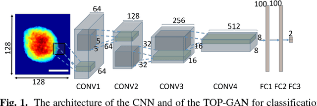
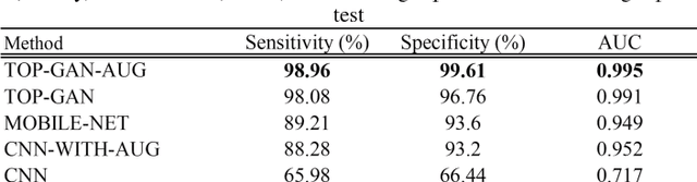
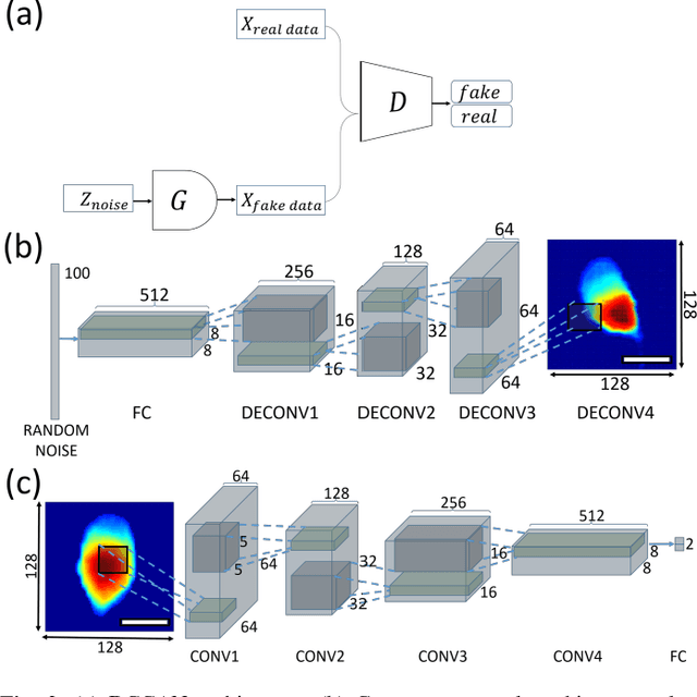
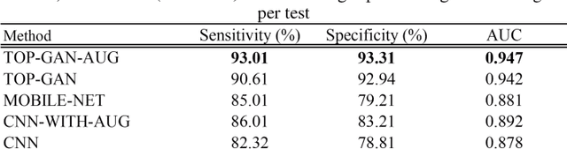
We propose a new deep learning approach for medical imaging that copes with the problem of a small training set, the main bottleneck of deep learning, and apply it for classification of healthy and cancer cells acquired by quantitative phase imaging. The proposed method, called transferring of pre-trained generative adversarial network (TOP-GAN), is a hybridization between transfer learning and generative adversarial networks (GANs). Healthy cells and cancer cells of different metastatic potential have been imaged by low-coherence off-axis holography. After the acquisition, the optical path delay maps of the cells have been extracted and directly used as an input to the deep networks. In order to cope with the small number of classified images, we have used GANs to train a large number of unclassified images from another cell type (sperm cells). After this preliminary training, and after transforming the last layer of the network with new ones, we have designed an automatic classifier for the correct cell type (healthy/primary cancer/metastatic cancer) with 90-99% accuracy, although small training sets of down to several images have been used. These results are better in comparison to other classic methods that aim at coping with the same problem of a small training set. We believe that our approach makes the combination of holographic microscopy and deep learning networks more accessible to the medical field by enabling a rapid, automatic and accurate classification in stain-free imaging flow cytometry. Furthermore, our approach is expected to be applicable to many other medical image classification tasks, suffering from a small training set.