 Add to Chrome
 Add to Firefox
 Add to Edge
Simultaneous Segmentation of Ventricles and Normal/Abnormal White Matter Hyperintensities in Clinical MRI using Deep Learning
Jun 08, 2025Multiple sclerosis (MS) diagnosis and monitoring rely heavily on accurate assessment of brain MRI biomarkers, particularly white matter hyperintensities (WMHs) and ventricular changes. Current segmentation approaches suffer from several limitations: they typically segment these structures independently despite their pathophysiological relationship, struggle to differentiate between normal and pathological hyperintensities, and are poorly optimized for anisotropic clinical MRI data. We propose a novel 2D pix2pix-based deep learning framework for simultaneous segmentation of ventricles and WMHs with the unique capability to distinguish between normal periventricular hyperintensities and pathological MS lesions. Our method was developed and validated on FLAIR MRI scans from 300 MS patients. Compared to established methods (SynthSeg, Atlas Matching, BIANCA, LST-LPA, LST-LGA, and WMH-SynthSeg), our approach achieved superior performance for both ventricle segmentation (Dice: 0.801+/-0.025, HD95: 18.46+/-7.1mm) and WMH segmentation (Dice: 0.624+/-0.061, precision: 0.755+/-0.161). Furthermore, our method successfully differentiated between normal and abnormal hyperintensities with a Dice coefficient of 0.647. Notably, our approach demonstrated exceptional computational efficiency, completing end-to-end processing in approximately 4 seconds per case, up to 36 times faster than baseline methods, while maintaining minimal resource requirements. This combination of improved accuracy, clinically relevant differentiation capability, and computational efficiency addresses critical limitations in current neuroimaging analysis, potentially enabling integration into routine clinical workflows and enhancing MS diagnosis and monitoring.
Characterization of Brain Cortical Morphology Using Localized Topology-Encoding Graphs
Oct 17, 2018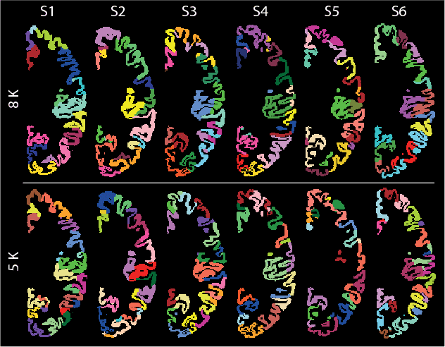
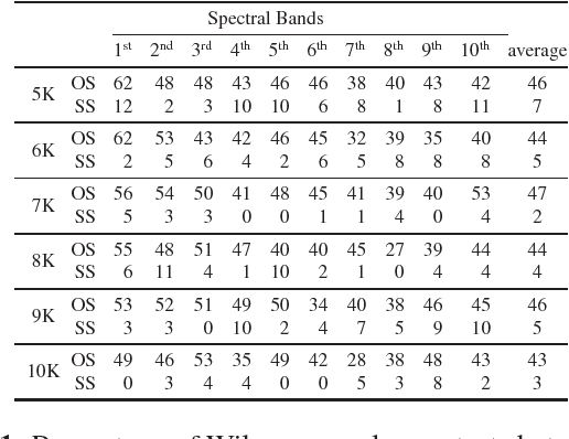
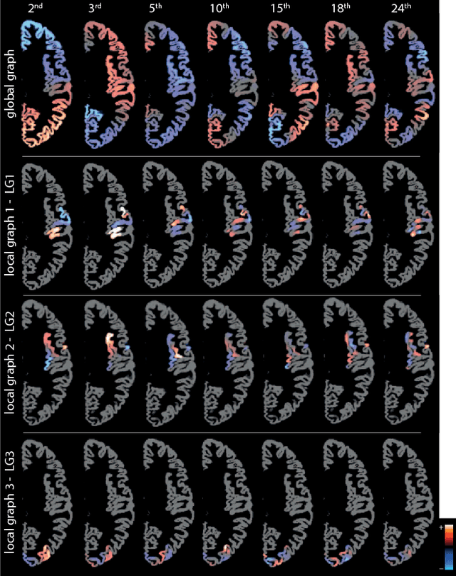
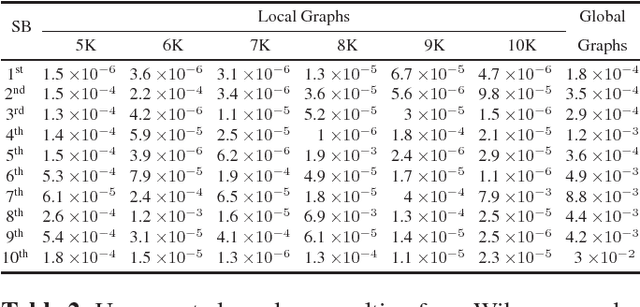
The human brain cortical layer has a convoluted morphology that is unique to each individual. Characterization of the cortical morphology is necessary in longitudinal studies of structural brain change, as well as in discriminating individuals in health and disease. A method for encoding the cortical morphology in the form of a graph is presented. The design of graphs that encode the global cerebral hemisphere cortices as well as localized cortical regions is proposed. Spectral features of these graphs are then studied and proposed as descriptors of cortical morphology. As proof-of-concept of their applicability in characterizing cortical morphology, the descriptors are studied in the context of discriminating individuals based on their sex.