 Add to Chrome
 Add to Firefox
 Add to Edge
Negative controls reveal volume-driven confounding in radiomics and imaging foundation model features
Jul 30, 2026
Radiomics and imaging foundation models promise non-invasive biomarkers of tumour biology, yet predictive signatures may reflect tumour volume or acquisition artifacts rather than meaningful image structure. We introduce READII-2-ROQC, an open-source framework that uses volume-preserving negative controls to assess whether radiomic and deep imaging features capture independent spatial signals. READII-2-ROQC generates voxel-perturbed images across tumour, background and whole-image regions using configurable randomization strategies, then compares feature behaviour and model performance between original and control images. Applied to three public cancer imaging cohorts, the framework processed 3,552 tumour volumes and extracted PyRadiomics and foundation-model features from original images and nine matched controls. Reproducing published survival and HPV-status signatures, we show that multiple models retain performance after spatial structure is destroyed, revealing volume-driven or contextual confounding, whereas others show perturbation-sensitive signal. READII-2-ROQC provides a scalable quality-control strategy for developing interpretable, biologically grounded imaging biomarkers and reproducible radiomics workflows.

A Machine Learning Challenge for Prognostic Modelling in Head and Neck Cancer Using Multi-modal Data
Jan 28, 2021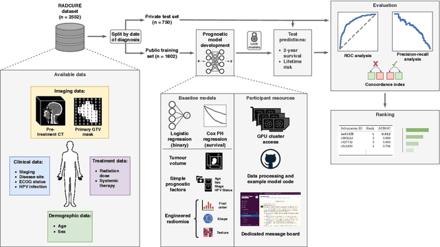
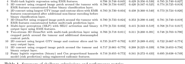
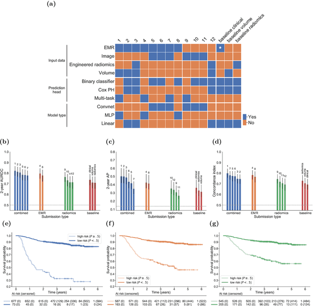
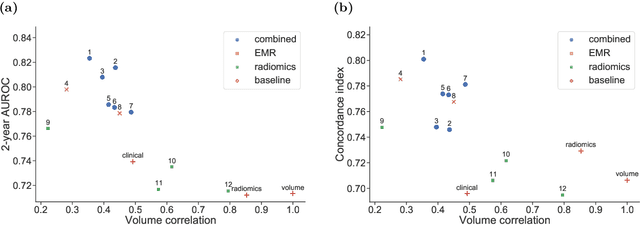
Accurate prognosis for an individual patient is a key component of precision oncology. Recent advances in machine learning have enabled the development of models using a wider range of data, including imaging. Radiomics aims to extract quantitative predictive and prognostic biomarkers from routine medical imaging, but evidence for computed tomography radiomics for prognosis remains inconclusive. We have conducted an institutional machine learning challenge to develop an accurate model for overall survival prediction in head and neck cancer using clinical data etxracted from electronic medical records and pre-treatment radiological images, as well as to evaluate the true added benefit of radiomics for head and neck cancer prognosis. Using a large, retrospective dataset of 2,552 patients and a rigorous evaluation framework, we compared 12 different submissions using imaging and clinical data, separately or in combination. The winning approach used non-linear, multitask learning on clinical data and tumour volume, achieving high prognostic accuracy for 2-year and lifetime survival prediction and outperforming models relying on clinical data only, engineered radiomics and deep learning. Combining all submissions in an ensemble model resulted in improved accuracy, with the highest gain from a image-based deep learning model. Our results show the potential of machine learning and simple, informative prognostic factors in combination with large datasets as a tool to guide personalized cancer care.