 Add to Chrome
 Add to Firefox
 Add to Edge
Mitosis Detection in the Wild: Multi-Tumor and Context-Aware Generalization in the MIDOG 2025 Challenge
Jun 05, 2026
Automated mitosis detection is a well-established task in computational pathology. While previous benchmarks focused on scanner-induced domain shift, clinical "real-world" application requires models to be robust across the vast variance to be expected in the histological landscape. The MItosis DOmain Generalization (MIDOG) 2025 challenge was designed to evaluate algorithmic performance across unprecedented biological and contextual diversity. We curated a test dataset of 365 cases, encompassing 12 distinct human, canine and feline tumor types, digitized across multiple scanning platforms. Moving beyond hand-selected hotspots, the challenge required detection also in random tissue areas (representative of the whole slide detection situation) and challenging areas (areas rich in hard negatives). In the second track, we introduced the classification of atypical mitotic figures (AMFs). There were 18 teams submitting to the detection track, with F1 scores ranging up to 0.740. In the AMF detection track, we had 21 submissions with balanced accuracy values up to 0.908. Our analysis reveals that while most models perform reliably in traditional hotspots, significant performance degradation occurs in challenging ROIs, where false positive rates tripled. Furthermore, performance varied significantly across the 12 tumor types, highlighting "blind spots" in current state-of-the-art architectures when encountering rare or highly pleomorphic malignancies. Moreover, we evaluated the effectiveness of ensembling and found a mean increases of 1.5 and 1.3 percentage points in F1 score and balanced accuracy, respectively. In contrast, TTA showed no relevant improvement. MIDOG 2025 demonstrates that "in the wild" mitosis detection remains a significant hurdle. The transition from hotspot-only evaluation to a multi-contextual framework provides a more realistic proxy for clinical reliability.

Vision Foundation Models in Agriculture: Toward Domain-Specific Adaptation for Weed Herbicide Trials Assessment
Nov 06, 2025Herbicide field trials require accurate identification of plant species and assessment of herbicide-induced damage across diverse environments. While general-purpose vision foundation models have shown promising results in complex visual domains, their performance can be limited in agriculture, where fine-grained distinctions between species and damage types are critical. In this work, we adapt a general-purpose vision foundation model to herbicide trial characterization. Trained using a self-supervised learning approach on a large, curated agricultural dataset, the model learns rich and transferable representations optimized for herbicide trials images. Our domain-specific model significantly outperforms the best general-purpose foundation model in both species identification (F1 score improvement from 0.91 to 0.94) and damage classification (from 0.26 to 0.33). Under unseen conditions (new locations and other time), it achieves even greater gains (species identification from 0.56 to 0.66; damage classification from 0.17 to 0.27). In domain-shift scenarios, such as drone imagery, it maintains strong performance (species classification from 0.49 to 0.60). Additionally, we show that domain-specific pretraining enhances segmentation accuracy, particularly in low-annotation regimes. An annotation-efficiency analysis reveals that, under unseen conditions, the domain-specific model achieves 5.4% higher F1 score than the general-purpose model, while using 80% fewer labeled samples. These results demonstrate the generalization capabilities of domain-specific foundation models and their potential to significantly reduce manual annotation efforts, offering a scalable and automated solution for herbicide trial analysis.
Unleashing the Potential of Synthetic Images: A Study on Histopathology Image Classification
Sep 24, 2024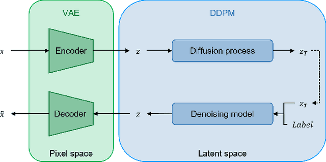
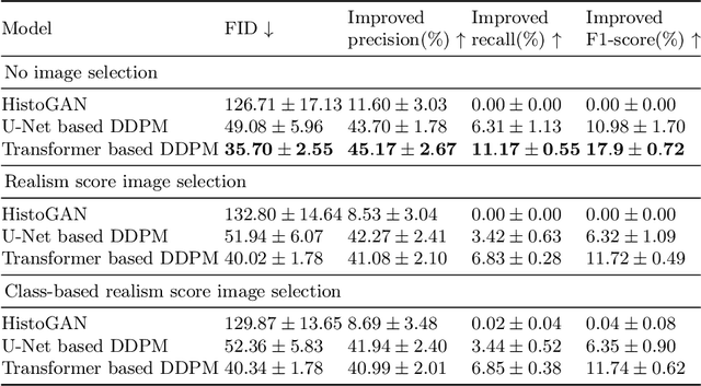
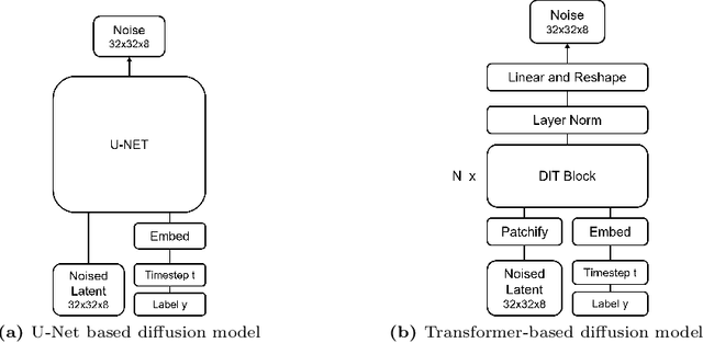
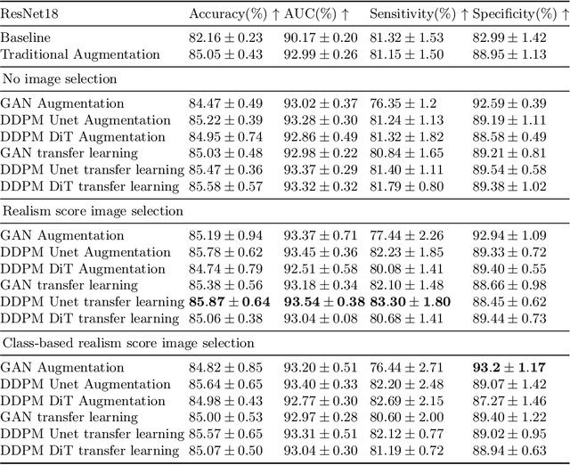
Histopathology image classification is crucial for the accurate identification and diagnosis of various diseases but requires large and diverse datasets. Obtaining such datasets, however, is often costly and time-consuming due to the need for expert annotations and ethical constraints. To address this, we examine the suitability of different generative models and image selection approaches to create realistic synthetic histopathology image patches conditioned on class labels. Our findings highlight the importance of selecting an appropriate generative model type and architecture to enhance performance. Our experiments over the PCam dataset show that diffusion models are effective for transfer learning, while GAN-generated samples are better suited for augmentation. Additionally, transformer-based generative models do not require image filtering, in contrast to those derived from Convolutional Neural Networks (CNNs), which benefit from realism score-based selection. Therefore, we show that synthetic images can effectively augment existing datasets, ultimately improving the performance of the downstream histopathology image classification task.