 Add to Chrome
 Add to Firefox
 Add to Edge
Denoising method for dynamic contrast-enhanced CT perfusion studies using three-dimensional deep image prior as a simultaneous spatial and temporal regularizer
Aug 31, 2022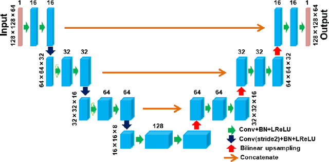
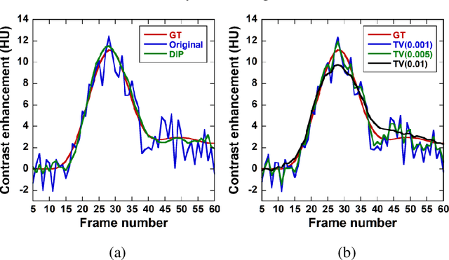
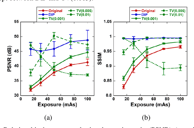
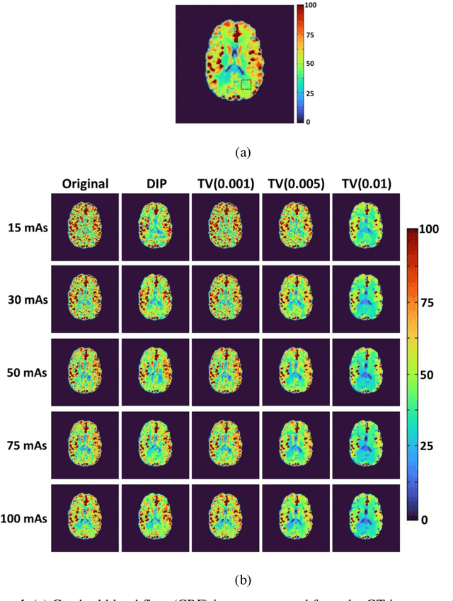
This study aimed to propose a denoising method for dynamic contrast-enhanced computed tomography (DCE-CT) perfusion studies using a three-dimensional deep image prior (DIP), and to investigate its usefulness in comparison with total variation (TV)-based methods with different regularization parameter (alpha) values through simulation studies. In the proposed DIP method, the DIP was incorporated into the constrained optimization problem for image denoising as a simultaneous spatial and temporal regularizer, which was solved using the alternating direction method of multipliers. In the simulation studies, DCE-CT images were generated using a digital brain phantom and their noise level was varied using the X-ray exposure noise model with different exposures (15, 30, 50, 75, and 100 mAs). Cerebral blood flow (CBF) images were generated from the original contrast enhancement (CE) images and those obtained by the DIP and TV methods using block-circulant singular value decomposition. The quality of the CE images was evaluated using the peak signal-to-noise ratio (PSNR) and structural similarity index (SSIM). To compare the CBF images obtained by the different methods and those generated from the ground truth images, linear regression analysis was performed. When using the DIP method, the PSNR and SSIM were not significantly dependent on the exposure, and the SSIM was the highest for all exposures. When using the TV methods, they were significantly dependent on the exposure and alpha values. The results of the linear regression analysis suggested that the linearity of the CBF images obtained by the DIP method was superior to those obtained from the original CE images and by the TV methods. Our preliminary results suggest that the DIP method is useful for denoising DCE-CT images at ultra-low to low exposures and for improving the accuracy of the CBF images generated from them.
Alternating direction method of multipliers applied to medical image restoration
Jul 04, 2021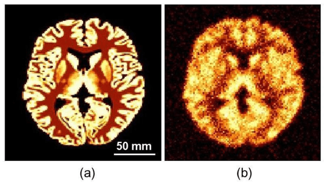
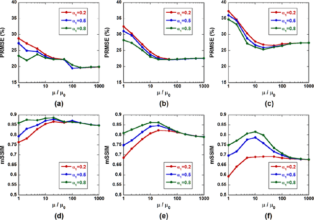
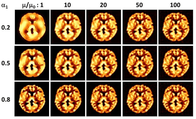
We investigate the effects of the regularization parameter for the norm () and penalty parameter () in the alternating direction method of multipliers (ADMM) on the quality of restored medical images. Simulation studies are performed using images degraded by a point spread function (PSF) and Gaussian noise. The j-th column of the system matrix () is calculated by convolving the image with unity at pixel j and zero at all other pixels and the PSF. The simulation studies show that the mean structural similarity index is maximal when is approximately 10 to 20, where , with and being the transpose of A and the observed data, respectively. The restored image became blurred with a decrease in . This study will be useful for identifying optimal parameter values in the ADMM when applied to medical image restoration.