 Add to Chrome
 Add to Firefox
 Add to Edge
X-ray phase and dark-field computed tomography without optical elements
Oct 14, 2023X-ray diffusive dark-field imaging, which allows spatially unresolved microstructure to be mapped across a sample, is an increasingly popular tool in an array of settings. Here, we present a new algorithm for phase and dark-field computed tomography based on the x-ray Fokker-Planck equation. Needing only a coherent x-ray source, sample, and detector, our propagation-based algorithm can map the sample density and dark-field/diffusion properties of the sample in 3D. Importantly, incorporating dark-field information in the density reconstruction process enables a higher spatial resolution reconstruction than possible with previous propagation-based approaches. Two sample exposures at each projection angle are sufficient for the successful reconstruction of both the sample density and dark-field Fokker-Planck diffusion coefficients. We anticipate that the proposed algorithm may be of benefit in biomedical imaging and industrial settings.
X-ray dark-field and phase retrieval without optics, via the Fokker-Planck equation
Dec 21, 2021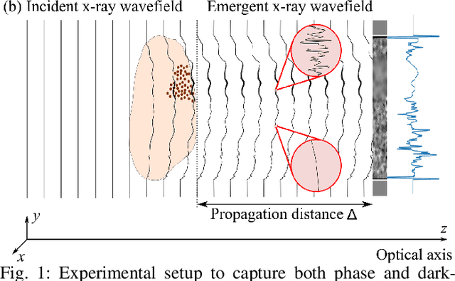
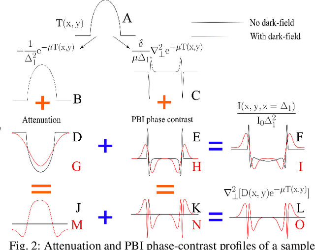
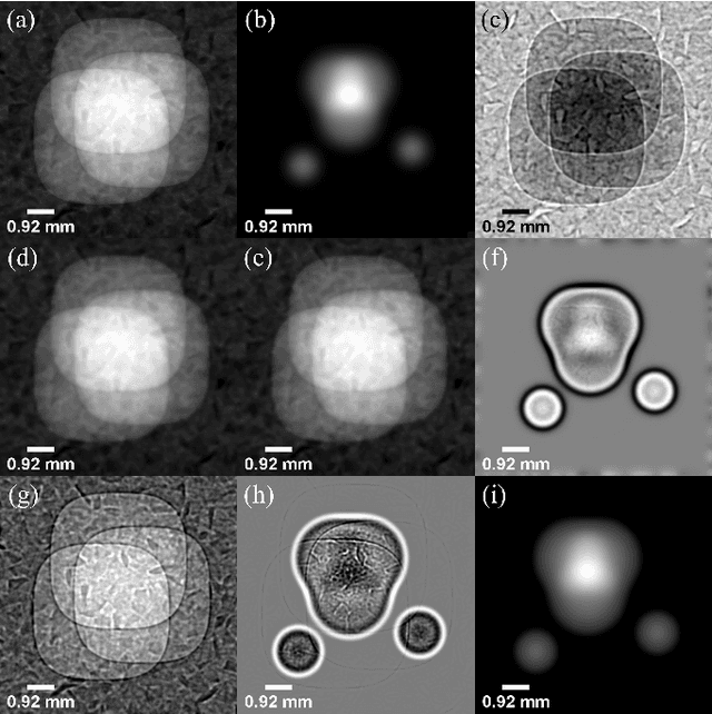
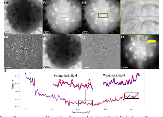
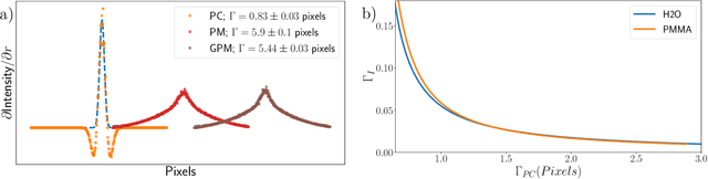
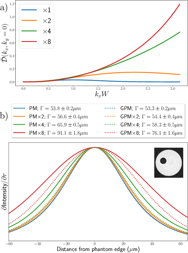
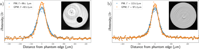
Emerging methods of x-ray imaging that capture phase and dark-field effects are equipping medicine with complementary sensitivity to conventional radiography. These methods are being applied over a wide range of scales, from virtual histology to clinical chest imaging, and typically require the introduction of optics such as gratings. Here, we consider extracting x-ray phase and dark-field signals from bright-field images collected using nothing more than a coherent x-ray source and detector. Our approach is based on the Fokker--Planck equation for paraxial imaging, which is the diffusive generalization of the transport-of-intensity equation. Specifically, we utilize the Fokker--Planck equation in the context of propagation-based phase-contrast imaging, where we show that two intensity images are sufficient for successful retrieval of the projected thickness and dark-field signals associated with the sample. We show the results of our algorithm using both a simulated dataset and an experimental dataset. These demonstrate that the x-ray dark-field signal can be extracted from propagation-based images, and that x-ray phase can be retrieved with better spatial resolution when dark-field effects are taken into account. We anticipate the proposed algorithm will be of benefit in biomedical imaging, industrial settings, and other non-invasive imaging applications.
Enhanced spatial resolution through DFT rederivations of X-ray phase retrieval algorithms
Jun 14, 2021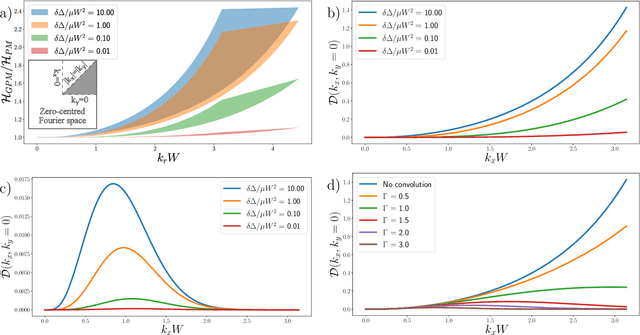
Propagation-based phase-contrast imaging, used in conjunction with the phase retrieval algorithm based on the Transport-of-Intensity Equation (TIE) (Paganin et al., 2002), is commonly used to improve the sensitivity of X-ray imaging. Recently, a `Generalised Paganin Method' algorithm was published to correct the tendency of the TIE algorithm to over-blur images. The article, Paganin et al. 2020, provided a derivation of the new method and demonstrated a difference in the level of blurring applied by each algorithm. In this manuscript, we quantify the spatial resolution improvement and describe the optimal experimental conditions to observe this improvement. We link the effectiveness of the spatial resolution improvement to the imaging point spread function (PSF), incorporating the PSF to compare the blurring applied by each algorithm. We then validate this model through measurements of spatial resolution in experimental data imaging plastic phantoms and biological tissue, using detectors with different PSFs. By analysing edge-spread functions in CT data captured with indirect detectors with PSFs of several pixels in extent, we show negligible spatial resolution improvement when using the generalised Paganin method. However, a clear improvement in spatial resolution, up to 17%, was observed with direct detectors having PSFs of approximately one pixel in extent. Additionally, we demonstrate clear visual improvement in resolution in CT slices of rat lungs. Finally, we demonstrate the versatility of this improvement by generalising other phase retrieval algorithms, namely for multi-material samples and for spectral decomposition using propagation-based phase contrast, and experimentally verify improvements in spatial resolution.