 Add to Chrome
 Add to Firefox
 Add to Edge
Disentangled Shared Representations Improve Morpho-Transcriptomic Integration
Aug 14, 2026
Spatial transcriptomics (ST) enables the simultaneous profiling of gene expression and tissue morphology, creating an opportunity to learn multimodal representations capturing shared morpho-transcriptomic structure. However, standard multimodal models often compress modalities into a common latent space without explicitly separating shared and modality-specific sources of variation, which may limit downstream utility. We investigate whether explicit disentanglement of shared and private latent components improves multimodal representation learning for paired Hematoxylin \& Eosin (H\&E) and ST data. We compare VAE-based and contrastive approaches, each in standard and disentangled variants, across two cancer cohorts under matched experimental conditions. Representations are evaluated using cross-modal reconstruction, downstream probing and cross-modal probe transfer. The experiments suggest two main trends. First, contrastive objectives yield higher downstream probing performance than VAE-based models. Second, disentangled variants improve the selected reconstruction and probing metrics, although the gains depend on the model family, task, direction, and disentanglement strength. Overall, our results suggest that explicitly factorizing shared and modality-specific information can improve multimodal representation learning for spatial transcriptomics and provides a useful evaluation framework for future foundation models.

Deep Learning model predicts the c-Kit-11 mutational status of canine cutaneous mast cell tumors by HE stained histological slides
Jan 02, 2024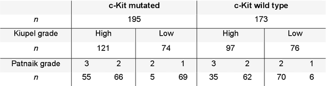
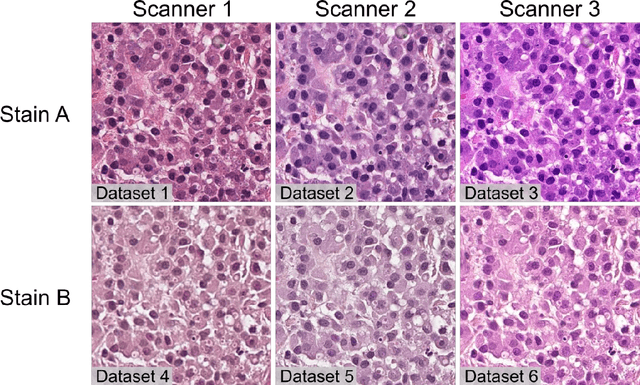
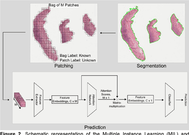
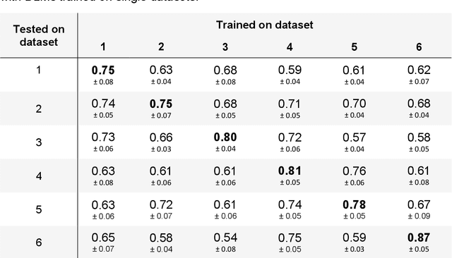
Numerous prognostic factors are currently assessed histopathologically in biopsies of canine mast cell tumors to evaluate clinical behavior. In addition, PCR analysis of the c-Kit exon 11 mutational status is often performed to evaluate the potential success of a tyrosine kinase inhibitor therapy. This project aimed at training deep learning models (DLMs) to identify the c-Kit-11 mutational status of MCTs solely based on morphology without additional molecular analysis. HE slides of 195 mutated and 173 non-mutated tumors were stained consecutively in two different laboratories and scanned with three different slide scanners. This resulted in six different datasets (stain-scanner variations) of whole slide images. DLMs were trained with single and mixed datasets and their performances was assessed under scanner and staining domain shifts. The DLMs correctly classified HE slides according to their c-Kit 11 mutation status in, on average, 87% of cases for the best-suited stain-scanner variant. A relevant performance drop could be observed when the stain-scanner combination of the training and test dataset differed. Multi-variant datasets improved the average accuracy but did not reach the maximum accuracy of algorithms trained and tested on the same stain-scanner variant. In summary, DLM-assisted morphological examination of MCTs can predict c-Kit-exon 11 mutational status of MCTs with high accuracy. However, the recognition performance is impeded by a change of scanner or staining protocol. Larger data sets with higher numbers of scans originating from different laboratories and scanners may lead to more robust DLMs to identify c-Kit mutations in HE slides.