 Add to Chrome
 Add to Firefox
 Add to Edge
SWITi: Quantifying and Reducing Tiling Artifacts with Sliding Window Inner Tiling
Jul 21, 2026
SWITi is a test-time method for reducing artifacts in tiled predictions, particularly for neural networks that learn posterior distributions from which solutions are sampled at inference time. Tiled predictions are unavoidable for large image data, and artifacts arise whenever tiles are smaller than a network's receptive field and when tiles are independent posterior samples. SWITi averages overlapping sliding-window predictions, so discrepancies between neighboring samples are spread across shifted tile positions rather than accumulating at fixed seam coordinates. For posterior models, SWITi uses no more tile samples than an MMSE estimate requires and therefore incurs no additional forward passes. Additionally, we introduce two reference-free metrics, the Fraction of Rejected Tests (FRT) and Artifact Severity (ASV), for detecting and quantifying tiling artifacts from a per-tile permutation test that compares the distribution of pixel gradients across tile seams against the surrounding image content. On pre-trained and published image splitting models across three fluorescence microscopy datasets in 2D and 3D, we show that SWITi substantially attenuates stitching seams while also improving reconstruction fidelity and resolution. Since tiling artifacts in posterior predictions can easily be mistaken for biological structures or for boundaries between biological structures, removing or reducing them using SWITi will improve the downstream processing of large image predictions, which is particularly relevant for biomedical data.

MicroSSIM: Improved Structural Similarity for Comparing Microscopy Data
Aug 16, 2024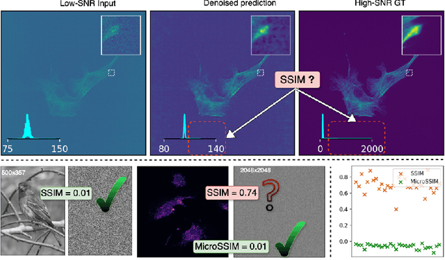
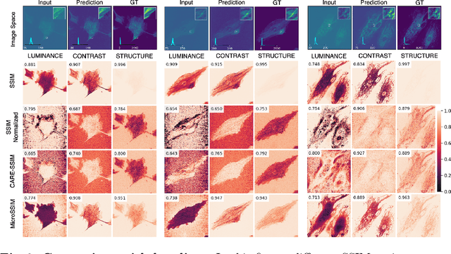
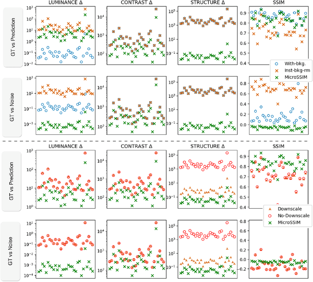
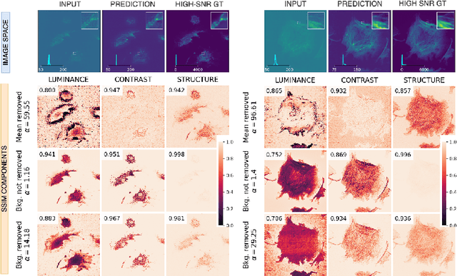
Microscopy is routinely used to image biological structures of interest. Due to imaging constraints, acquired images are typically low-SNR and contain noise. Over the last few years, regression-based tasks like unsupervised denoising and splitting have found utility in working with such noisy micrographs. For evaluation, Structural Similarity (SSIM) is one of the most popular measures used in the field. For such tasks, the best evaluation would be when both low-SNR noisy images and corresponding high-SNR clean images are obtained directly from a microscope. However, due to the following three peculiar properties of the microscopy data, we observe that SSIM is not well suited to this data regime: (a) high-SNR micrographs have higher intensity pixels as compared to low SNR micrographs, (b) high-SNR micrographs have higher intensity pixels than found in natural images, images for which SSIM was developed, and (c) a digitally configurable offset is added by the detector present inside the microscope. We show that SSIM components behave unexpectedly when the prediction generated from low-SNR input is compared with the corresponding high-SNR data. We explain this behavior by introducing the phenomenon of saturation, where the value of SSIM components becomes less sensitive to (dis)similarity between the images. We introduce microSSIM, a variant of SSIM, which overcomes the above-discussed issues. We justify the soundness and utility of microSSIM using theoretical and empirical arguments and show the utility of microSSIM on two tasks: unsupervised denoising and joint image splitting with unsupervised denoising. Since our formulation can be applied to a broad family of SSIM-based measures, we also introduce MicroMS3IM, a microscopy-specific variation of MS-SSIM. The source code and python package is available at https://github.com/juglab/MicroSSIM.
bAIoimage analysis: elevating the rate of scientific discovery -- as a community
Mar 29, 2023The future of bioimage analysis is increasingly defined by the development and use of tools that rely on deep learning and artificial intelligence (AI). For this trend to continue in a way most useful for stimulating scientific progress, it will require our multidisciplinary community to work together, establish FAIR data sharing and deliver usable, reproducible analytical tools.