 Add to Chrome
 Add to Firefox
 Add to Edge
Far-field compressive ultrasound beamforming
Mar 23, 2026We present a compressive beamforming method for coherent plane-wave compounding (CPWC) ultrasound imaging based on a far-field decomposition of the received radiofrequency (RF) data into virtual plane waves. This decomposition recasts the imaging operation entirely in the spatial frequency domain ($k$-space), allowing direct and flexible control over $k$-space sampling distributions based on the principle of coarrays. We present vernier-type sampling strategies designed to optimize the tradeoff between image contrast and resolution with minimum redundancy, including strategies that favor dense low-frequency sampling for high contrast, shifted schemes that extend the frequency support for improved resolution, and confocal or hybrid compounding schemes that approximate the spatial-frequency transfer function of conventional DAS beamforming. Our method, called KK beamforming, is validated with a calibration phantom and in-vivo human tissue data, demonstrating compression factors of an order of magnitude while maintaining image qualities comparable to conventional DAS. We further demonstrate that KK beamforming yields improvements in computational speed owing to its reduced memory footprint and more efficient cache utilization of the compressed data and associated look-up tables.
Plane-wave compounding with adaptive joint coherence factor weighting
Apr 18, 2024


Coherent Plane Wave Compounding (CPWC) is widely used for ultrasound imaging. This technique involves sending plane waves into a sample at different transmit angles and recording the resultant backscattered echo at different receive positions. The time-delayed signals from the different combinations of transmit angles and receive positions are then coherently summed to produce a beamformed image. Various techniques have been developed to characterize the quality of CPWC beamforming based on the measured coherence across the transmit or receive apertures. Here, we propose a more fine-grained approach where the signals from every transmit/receive combination are separately evaluated using a quality metric based on their joint spatio-angular coherence. The signals are then individually weighted according to their measured Joint Coherence Factor (JCF) prior to being coherently summed. To facilitate the comparison of JCF beamforming compared to alternative techniques, we further propose a method of image display standardization based on contrast matching. We show results from tissue-mimicking phantoms and human soft-tissue imaging. Fine-grained JCF weighting is found to improve CPWC image quality compared to alternative approaches.
Interferometric speckle visibility spectroscopy (iSVS) for measuring decorrelation time and dynamics of moving samples with enhanced signal-to-noise ratio and relaxed reference requirements
Jun 30, 2023


Diffusing wave spectroscopy (DWS) is a group of techniques used to measure the dynamics of a scattering medium in a non-invasive manner. DWS methods rely on detecting the speckle light field from the moving scattering media and measuring the speckle decorrelation time to quantify the scattering mediums dynamics. For DWS, the signal-to-noise (SNR) is determined by the ratio between measured decorrelation time to the standard error of the measurement. This SNR is often low in certain applications because of high noise variances and low signal intensity, especially in biological applications with restricted exposure and emission levels. To address this photon-limited signal-to-noise ratio problem, we investigated, theoretically and experimentally, the SNR of an interferometric speckle visibility spectroscopy (iSVS) compared to more traditional DWS methods. We found that iSVS can provide excellent SNR performance through its ability to overcome camera noise. We also proved iSVS system has more relaxed constraints on the reference beam properties than most other interferometric systems. For an iSVS to function properly, we simply require the reference beam to exhibit local temporal stability, while incident angle, reference phase, and intensity uniformity do not need to be constrained. This flexibility can potentially enable more unconventional iSVS implementation schemes.
Ultrasound differential phase contrast using backscattering and the memory effect
Mar 14, 2021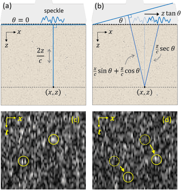
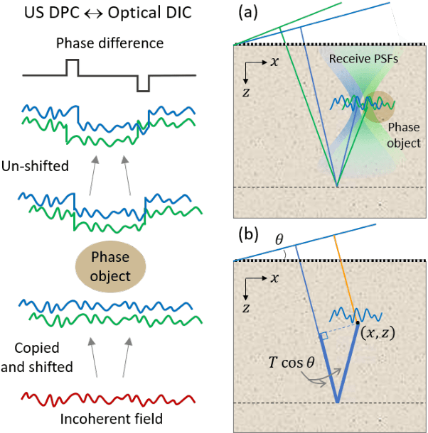
We describe a simple and fast technique to perform ultrasound differential phase contrast (DPC) imaging in arbitrarily thick scattering media. Though configured in a reflection geometry, DPC is based on transmission imaging and is a direct analogue of optical differential interference contrast (DIC). DPC exploits the memory effect and works in combination with standard pulse-echo imaging, with no additional hardware or data requirements, enabling complementary phase contrast (in the transverse direction) without any need for intensive numerical computation. We experimentally demonstrate the principle of DPC using tissue phantoms with calibrated speed-of-sound inclusions.