 Add to Chrome
 Add to Firefox
 Add to Edge
Performance Validation of Coded Wavefront Sensing for Quantitative Phase Imaging of Static and Dynamic Specimens Using Digital Holographic Microscopy
Aug 23, 2025Coded wavefront sensing (Coded-WFS) is a snapshot quantitative phase imaging (QPI) technique that has been shown to successfully leverage the memory effect to retrieve the phase of biological specimens. In this paper, we perform QPI on static silica beads and dynamic HEK cells using Coded-WFS. The accuracy of the retrieved phase map is validated using digital holographic microscopy (DHM) for the same specimens. We report comparisons of simultaneous bright-field intensity and optical path delay.
Direct Image Classification from Fourier Ptychographic Microscopy Measurements without Reconstruction
May 08, 2025The computational imaging technique of Fourier Ptychographic Microscopy (FPM) enables high-resolution imaging with a wide field of view and can serve as an extremely valuable tool, e.g. in the classification of cells in medical applications. However, reconstructing a high-resolution image from tens or even hundreds of measurements is computationally expensive, particularly for a wide field of view. Therefore, in this paper, we investigate the idea of classifying the image content in the FPM measurements directly without performing a reconstruction step first. We show that Convolutional Neural Networks (CNN) can extract meaningful information from measurement sequences, significantly outperforming the classification on a single band-limited image (up to 12 %) while being significantly more efficient than a reconstruction of a high-resolution image. Furthermore, we demonstrate that a learned multiplexing of several raw measurements allows maintaining the classification accuracy while reducing the amount of data (and consequently also the acquisition time) significantly.
Light-field Microscopy with a Consumer Light-field Camera
Dec 07, 2015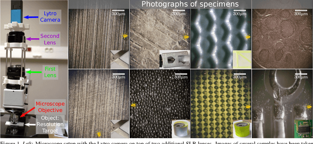
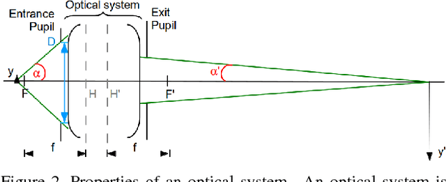
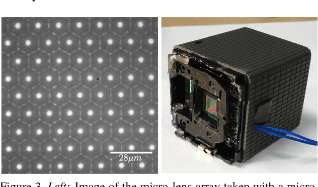
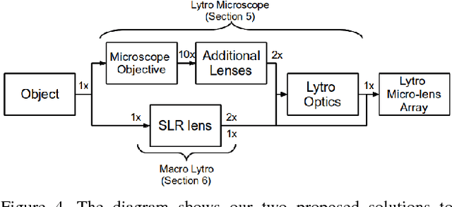
We explore the use of inexpensive consumer light- field camera technology for the purpose of light-field mi- croscopy. Our experiments are based on the Lytro (first gen- eration) camera. Unfortunately, the optical systems of the Lytro and those of microscopes are not compatible, lead- ing to a loss of light-field information due to angular and spatial vignetting when directly recording microscopic pic- tures. We therefore consider an adaptation of the Lytro op- tical system. We demonstrate that using the Lytro directly as an oc- ular replacement, leads to unacceptable spatial vignetting. However, we also found a setting that allows the use of the Lytro camera in a virtual imaging mode which prevents the information loss to a large extent. We analyze the new vir- tual imaging mode and use it in two different setups for im- plementing light-field microscopy using a Lytro camera. As a practical result, we show that the camera can be used for low magnification work, as e.g. common in quality control, surface characterization, etc. We achieve a maximum spa- tial resolution of about 6.25{\mu}m, albeit at a limited SNR for the side views.