 Add to Chrome
 Add to Firefox
 Add to Edge
Local Adaptation Improves Accuracy of Deep Learning Model for Automated X-Ray Thoracic Disease Detection : A Thai Study
May 12, 2020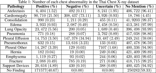
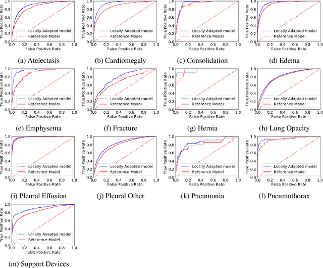
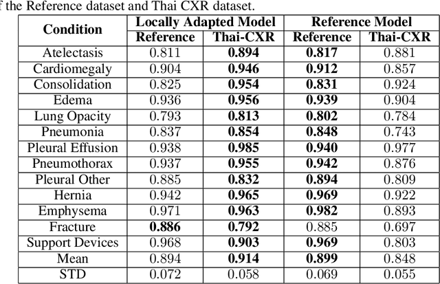
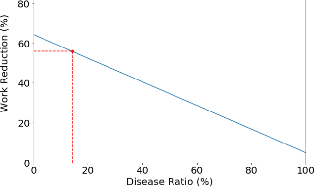
Despite much promising research in the area of artificial intelligence for medical image diagnosis, there has been no large-scale validation study done in Thailand to confirm the accuracy and utility of such algorithms when applied to local datasets. Here we present a wide-reaching development and testing of a deep learning algorithm for automated thoracic disease detection, utilizing 421,859 local chest radiographs. Our study shows that convolutional neural networks can achieve remarkable performance in detecting 13 common abnormality conditions on chest X-ray, and the incorporation of local images into the training set is key to the model's success. This paper presents a state-of-the-art model for CXR abnormality detection, reaching an average AUROC of 0.91. This model, if integrated to the workflow, can result in up to 55.6% work reduction for medical practitioners in the CXR analysis process. Our work emphasizes the importance of investing in local research of medical diagnosis algorithms to ensure safe and efficient usage within the intended region.
Automated Cardiothoracic Ratio Calculation and Cardiomegaly Detection using Deep Learning Approach
Feb 18, 2020
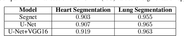
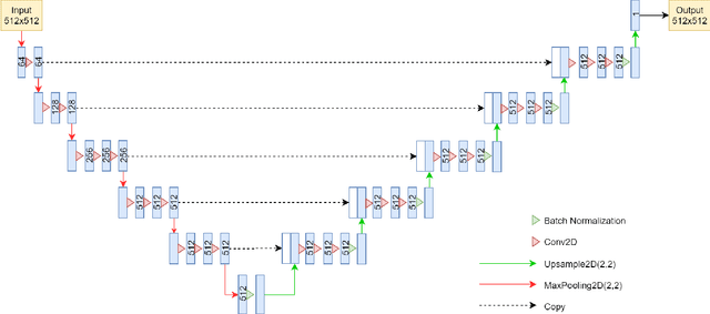
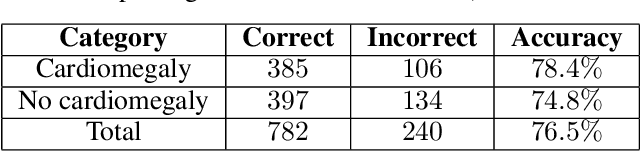
We propose an algorithm for calculating the cardiothoracic ratio (CTR) from chest X-ray films. Our approach applies a deep learning model based on U-Net with VGG16 encoder to extract lung and heart masks from chest X-ray images and calculate CTR from the extents of obtained masks. Human radiologists evaluated our CTR measurements, and $76.5\%$ were accepted to be included in medical reports without any need for adjustment. This result translates to a large amount of time and labor saved for radiologists using our automated tools.