 Add to Chrome
 Add to Firefox
 Add to Edge
Advancing Brain-Machine Interfaces: High Data Rate Battery-Free Implants
Jul 31, 2026
Implantable wireless brain-machine interfaces (BMI) encounter significant challenges in miniaturization, power consumption, and high data volume. While systems utilizing high resolution microelectrode arrays offer precision brain readout and/or stimulation, achieving high-rate wireless connectivity (32-128 Mbps) consumes excessive power, unsuitable for long-term use with implant batteries. This paper addresses wireless connectivity and power challenges by employing radio frequency backscatter and near-field wireless charging. This approach eliminates transceiver electronics in the implantable, reducing implant power consumption by offloading complexity to off-body reader electronics. It enables wireless powering of implantable neural recording and stimulation chips through magnetic coupling, enabling a fully implantable brain-machine interface. We present preliminary test results for this design scenario, demonstrating the feasibility of our approach.
* 3 pages, 4 figures. Supported by B-CRATOS project, Horizon 2020 FET-OPEN, grant 965044

Safety Analysis of Metasurface-Based Near-field Wireless Power Transfer System for Deep Implant
Jul 31, 2026Wireless power transfer is a method for energizing future implantable medical electronics. In this study, a metasurface-based near-field magnetic wireless power transfer system for deep implants is presented, and electromagnetic safety parameters, including field distributions, specific absorption rate (SAR), and temperature variations, are evaluated. The power transfer is modeled for a receiver implant at a distance of 8.5 cm from the designed metasurface. Based on the results, a maximum localized SAR of 0.072 mW/kg is achieved when the efficiency is 1.62%. Moreover, continuous power transfer shows that the local tissue temperature rises by less than 1.1 degrees Celsius.
* 5 pages, 7 figures, 3 tables. Funded by Research Council of Norway (grant 287112), South-Eastern Norway Regional Health Authority (grant 2020049), and B-CRATOS Horizon 2020 FET-OPEN (grant 965044)
Frequency demodulation with magnetoelectric coreshells: A novel approach to enhanced bio-stimulation
Jul 31, 2026Magnetoelectric (ME) coreshell devices have interesting applications in biomedical technologies including biosensing and communication, due to their strong inter-coupling between the magnetostrictive core and the piezoelectric shell. This property could be utilized for specific applications in localized bio-stimulation of cells. This paper provides a conceptual proof of using the non-linear property of ME coreshell devices for frequency demodulation and stimulation. We use the Multiphysics simulation approach, wherein the ME coreshell was biased with a DC magnetic field and perturbed through dual coil alternating magnetic fields. The combined effect of the ME non-linear magnetostriction and the dual coil perturbation resulted in the demodulation of the interference frequency component, that induced equivalent electrical hotspots on the piezoelectric shell. We provide a cross model verification, where the generated electrical current density on the piezo shell was provided as an input to a Hodgkin-Huxley (HH) neural cell model to actively induce membrane potentials on the cell. Future applications as a standalone, battery- and electronics-free, controllable, multi-functional and localized coreshells for targeted drug delivery, backscatter communication and bio-stimulation, is envisioned.
* 6 pages, 7 figures, 1 table. CC BY 4.0. Supported by B-CRATOS project, Horizon 2020 FET-OPEN, grant 965044, and ERCIM Alain Bensoussan Fellowship Programme
Propagation models for IEEE 802.15.6 standardization of implant communication in body area networks
Jul 24, 2026A body area network is a radio communication protocol for short-range, low-power, and highly reliable wireless communication for use on the surface, inside, or in the peripheral proximity of the human body. Combined with various biomedical sensors, BANs enable realtime collection and monitoring of physiological signals. Therefore, it is regarded as an important technology for the treatment and prevention of chronic diseases, and health monitoring of the elderly. The IEEE 802 LAN/MAN Standards Committee approved Task Group TG15.6 in December 2007. As a result of more than four years of effort, in February 2012, TG15.6 published the first international standard for BANs, IEEE Std 802.15.6. Throughout the development of this standard, ample collaboration between the standardization group and the research community was required. In particular, understanding the radio propagation mechanisms for BANs demanded the most research effort. Technical challenges were magnified for the case of implant communication because of the impossibility of conducting in-body measurements with human subjects. Therefore, research in this field had to make use of intricate computer simulations. This article outlines some of the research that has been done to obtain accurate propagation models supporting the standardization of implant communication in BANs. Current research to enhance the channel models of IEEE Std 802.15.6 through the use of ultra wideband signals for implantable devices along with physical measurements in animals is also presented.
Ultra-wideband statistical propagation channel model for implant sensors in the human chest
Jul 24, 2026Implant medical wireless sensors for monitoring physiological parameters, automatic drug provision, and so on represent a new promising healthcare technology. Inherent characteristics of ultra-wideband (UWB) radio make this technology highly suitable for the wireless interface of implant sensors. A communication channel model is essential for developing these wireless systems. However, there are currently few models describing the radio propagation inside the human body. To address this problem, a statistical model is presented for UWB propagation channels inside the human chest in the 1-6 GHz frequency range. The proposed statistical model is developed from numerical simulations using a heterogeneous anatomical model that includes the frequency-dependent dielectric properties of different human tissues. Mathematical formulas for the computation of path loss, scattering and the statistical implementation of the channel impulse response at different depths inside the chest are described. Two typical depths for implanted sensors in the chest, namely 20 and 80 mm are analysed in detail. Average path loss of approximately 20 and 50 dB is observed in each case, respectively. Moreover, the channel exhibits little time dispersion with a root-mean-square delay spread below 1 ns in both cases. These results aim at facilitating the tasks associated with the design of in-body medical communication systems.
* 8 pages, 9 figures, 1 table. Supported by the MELODY Project, Research Council of Norway (contract no. 187857/S10)
Antenna System for Simultaneous Wireless Power and Information Transfer to Brain Implants
Jul 02, 2026Brain-Computer Interfaces (BCIs) have revolutionized neuroscience applications, from motor rehabilitation to neuroergonomics. Traditional implantable BCIs with invasive microelectrode arrays pose challenges, notably the need for wired connections and inherent implantation risks. This paper introduces a battery-free wireless BCI system, consolidating an implant and its external supporting system. Our design centers on a dual-function antenna system: firstly, an inductive coupling mechanism enables wireless power transfer, sufficiently powering the implant's Application-Specific Integrated Circuit (ASIC) for stimulation and readout without an implant battery. Secondly, a backscatter antenna in the implant facilitates battery-free, high-data-rate wireless connectivity (up to 32 Mbps). This system not only enhances the BCI experience by eliminating wires but also retains data fidelity and energy efficiency, promising a safer, more efficient interface for tasks like robotic arm control.
* Published at IMBioC 2024. Part of the B-CRATOS project, Horizon 2020 FET-OPEN, grant 965044
Deep Learning for Improved Polyp Detection from Synthetic Narrow-Band Imaging
Jan 24, 2024To cope with the growing prevalence of colorectal cancer (CRC), screening programs for polyp detection and removal have proven their usefulness. Colonoscopy is considered the best-performing procedure for CRC screening. To ease the examination, deep learning based methods for automatic polyp detection have been developed for conventional white-light imaging (WLI). Compared with WLI, narrow-band imaging (NBI) can improve polyp classification during colonoscopy but requires special equipment. We propose a CycleGAN-based framework to convert images captured with regular WLI to synthetic NBI (SNBI) as a pre-processing method for improving object detection on WLI when NBI is unavailable. This paper first shows that better results for polyp detection can be achieved on NBI compared to a relatively similar dataset of WLI. Secondly, experimental results demonstrate that our proposed modality translation can achieve improved polyp detection on SNBI images generated from WLI compared to the original WLI. This is because our WLI-to-SNBI translation model can enhance the observation of polyp surface patterns in the generated SNBI images.
Multimodal Deep Learning for Personalized Renal Cell Carcinoma Prognosis: Integrating CT Imaging and Clinical Data
Jul 07, 2023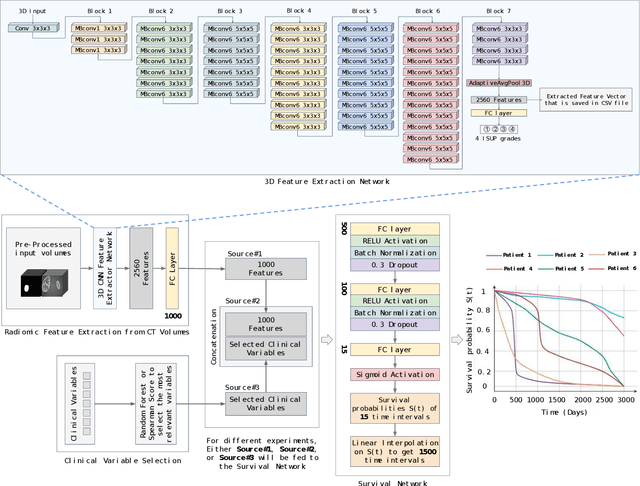
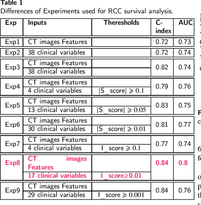
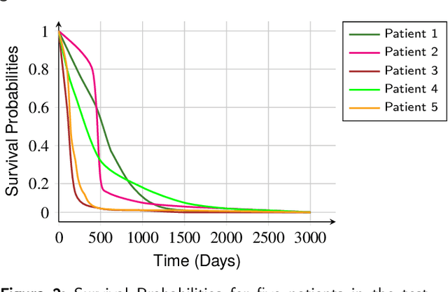

Renal cell carcinoma represents a significant global health challenge with a low survival rate. This research aimed to devise a comprehensive deep-learning model capable of predicting survival probabilities in patients with renal cell carcinoma by integrating CT imaging and clinical data and addressing the limitations observed in prior studies. The aim is to facilitate the identification of patients requiring urgent treatment. The proposed framework comprises three modules: a 3D image feature extractor, clinical variable selection, and survival prediction. The feature extractor module, based on the 3D CNN architecture, predicts the ISUP grade of renal cell carcinoma tumors linked to mortality rates from CT images. A selection of clinical variables is systematically chosen using the Spearman score and random forest importance score as criteria. A deep learning-based network, trained with discrete LogisticHazard-based loss, performs the survival prediction. Nine distinct experiments are performed, with varying numbers of clinical variables determined by different thresholds of the Spearman and importance scores. Our findings demonstrate that the proposed strategy surpasses the current literature on renal cancer prognosis based on CT scans and clinical factors. The best-performing experiment yielded a concordance index of 0.84 and an area under the curve value of 0.8 on the test cohort, which suggests strong predictive power. The multimodal deep-learning approach developed in this study shows promising results in estimating survival probabilities for renal cell carcinoma patients using CT imaging and clinical data. This may have potential implications in identifying patients who require urgent treatment, potentially improving patient outcomes. The code created for this project is available for the public on: \href{https://github.com/Balasingham-AI-Group/Survival_CTplusClinical}{GitHub}
Accurate Real-time Polyp Detection in Videos from Concatenation of Latent Features Extracted from Consecutive Frames
Mar 10, 2023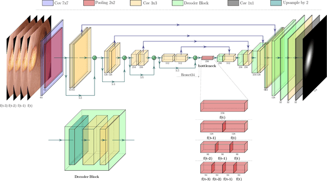
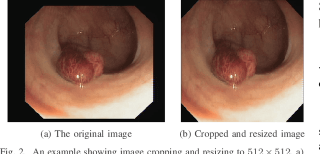
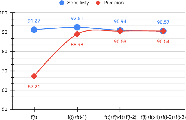
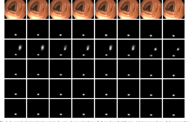
An efficient deep learning model that can be implemented in real-time for polyp detection is crucial to reducing polyp miss-rate during screening procedures. Convolutional neural networks (CNNs) are vulnerable to small changes in the input image. A CNN-based model may miss the same polyp appearing in a series of consecutive frames and produce unsubtle detection output due to changes in camera pose, lighting condition, light reflection, etc. In this study, we attempt to tackle this problem by integrating temporal information among neighboring frames. We propose an efficient feature concatenation method for a CNN-based encoder-decoder model without adding complexity to the model. The proposed method incorporates extracted feature maps of previous frames to detect polyps in the current frame. The experimental results demonstrate that the proposed method of feature concatenation improves the overall performance of automatic polyp detection in videos. The following results are obtained on a public video dataset: sensitivity 90.94\%, precision 90.53\%, and specificity 92.46%
Non-invasive Waveform Analysis for Emergency Triage via Simulated Hemorrhage: An Experimental Study using Novel Dynamic Lower Body Negative Pressure Model
Mar 01, 2023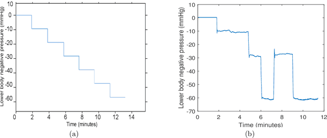
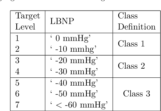
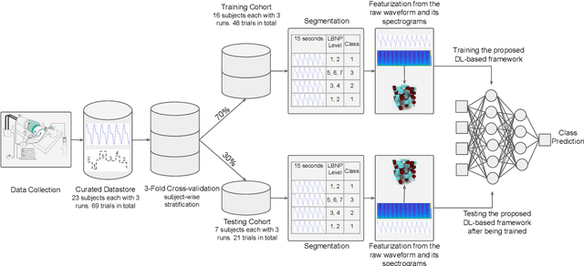
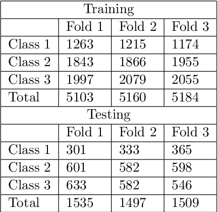
The extent to which advanced waveform analysis of non-invasive physiological signals can diagnose levels of hypovolemia remains insufficiently explored. The present study explores the discriminative ability of a deep learning (DL) framework to classify levels of ongoing hypovolemia, simulated via novel dynamic lower body negative pressure (LBNP) model among healthy volunteers. We used a dynamic LBNP protocol as opposed to the traditional model, where LBNP is applied in a predictable step-wise, progressively descending manner. This dynamic LBNP version assists in circumventing the problem posed in terms of time dependency, as in real-life pre-hospital settings, intravascular blood volume may fluctuate due to volume resuscitation. A supervised DL-based framework for ternary classification was realized by segmenting the underlying noninvasive signal and labeling segments with corresponding LBNP target levels. The proposed DL model with two inputs was trained with respective time-frequency representations extracted on waveform segments to classify each of them into blood volume loss: Class 1 (mild); Class 2 (moderate); or Class 3 (severe). At the outset, the latent space derived at the end of the DL model via late fusion among both inputs assists in enhanced classification performance. When evaluated in a 3-fold cross-validation setup with stratified subjects, the experimental findings demonstrated PPG to be a potential surrogate for variations in blood volume with average classification performance, AUROC: 0.8861, AUPRC: 0.8141, $F1$-score:72.16%, Sensitivity:79.06 %, and Specificity:89.21 %. Our proposed DL algorithm on PPG signal demonstrates the possibility of capturing the complex interplay in physiological responses related to both bleeding and fluid resuscitation using this challenging LBNP setup.