 Add to Chrome
 Add to Firefox
 Add to Edge
Single-shot refractive index slice imaging using spectrally multiplexed optical transfer function reshaping
Jan 13, 2023


The refractive index (RI) of cells and tissues is crucial in pathophysiology as a noninvasive and quantitative imaging contrast. Although its measurements have been demonstrated using three-dimensional quantitative phase imaging methods, these methods often require bulky interferometric setups or multiple measurements, which limits the measurement sensitivity and speed. Here, we present a single-shot RI imaging method that visualizes the RI of the in-focus region of a sample. By exploiting spectral multiplexing and optical transfer function engineering, three color-coded intensity images of a sample with three optimized illuminations were simultaneously obtained in a single-shot measurement. The measured intensity images were then deconvoluted to obtain the RI image of the in-focus slice of the sample. As a proof of concept, a setup was built using Fresnel lenses and a liquid-crystal display. For validation purposes, we measured microspheres of known RI and cross-validated the results with simulated results. Various static and highly dynamic biological cells were imaged to demonstrate that the proposed method can conduct single-shot RI slice imaging of biological samples with subcellular resolution.
Quantitative phase and refractive index imaging of 3D objects via optical transfer function reshaping
Jan 21, 2022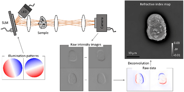
Deconvolution phase microscopy enables high-contrast visualization of transparent samples through reconstructions of their transmitted phases or refractive indexes. Herein, we propose a method to extend 2D deconvolution phase microscopy to thick 3D samples. The refractive index distribution of a sample can be obtained at a specific axial plane by measuring only four intensity images obtained under optimized illumination patterns. Also, the optical phase delay of a sample can be measured using different illumination patterns.