 Add to Chrome
 Add to Firefox
 Add to Edge
A microwave super-resolution imaging approach towards breast cancer margin mapping
Apr 23, 2026Accurate characterisation of margins in excised breast cancer tumours is critical to the success of surgical interventions, yet margin status is typically confirmed post-operatively using histopathology. Here we present a new approach to intraoperative margin assessment based on microwave single pixel imaging, demonstrating tissue phantom hydration mapping across large areas (~10 cm x 10 cm) at ~1 mm resolution. By leveraging the photo-induced change in microwave transparency of a silicon modulator placed under the sample, we map the microwave reflectivity and identify positive margins with deeply sub-wavelength resolution. We test the discriminatory capabilities of our approach using gelatine-based tumour phantoms with variations in water density representative of the margin and cancerous tissues of a resected tumour. We demonstrate the capability to identify, locate and quantify inadequate margins up to the typically targeted minimum thickness of 2 mm. Furthermore, using numerical modelling, we show that our approach is expected to be resilient to patient-specific tissue differences. Our technique has potential for future deployment as a real-time intraoperative tissue margin analysis tool.
Tracking moving objects through scattering media via speckle correlations
Feb 22, 2022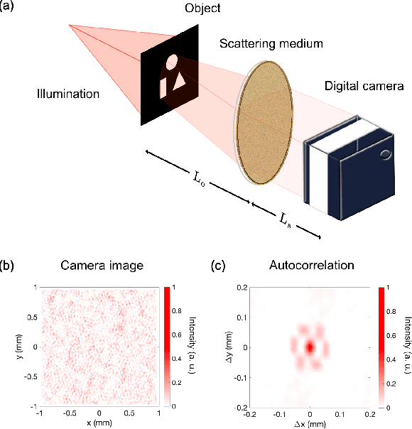
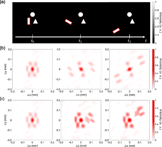
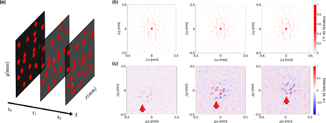
Scattering can rapidly degrade our ability to form an optical image, to the point where only speckle-like patterns can be measured. Truly non-invasive imaging through a strongly scattering obstacle is difficult, and usually reliant on a computationally intensive numerical reconstruction. In this work we show that, by combining the cross-correlations of the measured speckle pattern at different times, it is possible to track a moving object with minimal computational effort and over a large field of view.
Ghost Image Processing
Dec 14, 2021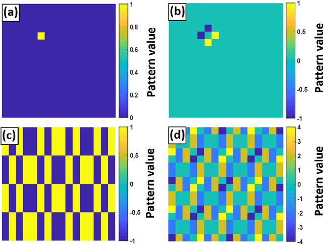
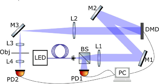
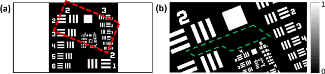
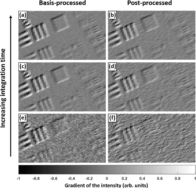
In computational ghost imaging the object is illuminated with a sequence of known patterns, and the scattered light is collected using a detector that has no spatial resolution. Using those patterns and the total intensity measurement from the detector, one can reconstruct the desired image. Here we study how the reconstructed image is modified if the patterns used for the reconstruction are not the same as the illumination patterns, and show that one can choose how to illuminate the object, such that the reconstruction process behaves like a spatial filtering operation on the image. The ability to measure directly a processed image, allows one to bypass the post-processing steps, and thus avoid any noise amplification they imply. As a simple example we show the case of an edge-detection filter.