 Add to Chrome
 Add to Firefox
 Add to Edge
Automatic Quantification of Volumes and Biventricular Function in Cardiac Resonance. Validation of a New Artificial Intelligence Approach
Jun 03, 2022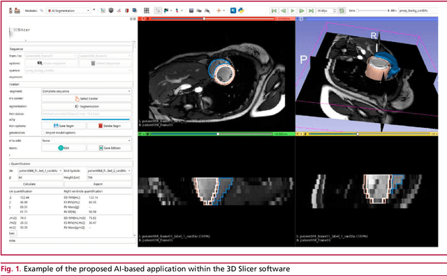
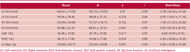
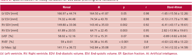
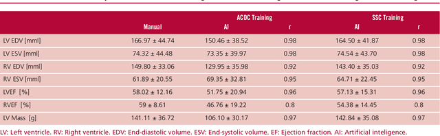
Background: Artificial intelligence techniques have shown great potential in cardiology, especially in quantifying cardiac biventricular function, volume, mass, and ejection fraction (EF). However, its use in clinical practice is not straightforward due to its poor reproducibility with cases from daily practice, among other reasons. Objectives: To validate a new artificial intelligence tool in order to quantify the cardiac biventricular function (volume, mass, and EF). To analyze its robustness in the clinical area, and the computational times compared with conventional methods. Methods: A total of 189 patients were analyzed: 89 from a regional center and 100 from a public center. The method proposes two convolutional networks that include anatomical information of the heart to reduce classification errors. Results: A high concordance (Pearson coefficient) was observed between manual quantification and the proposed quantification of cardiac function (0.98, 0.92, 0.96 and 0.8 for volumes and biventricular EF) in about 5 seconds per study. Conclusions: This method quantifies biventricular function and volumes in seconds with an accuracy equivalent to that of a specialist.
Detection of Fibrosis in Cine Magnetic Resonance Images Using Artificial Intelligence Techniques
Jun 03, 2022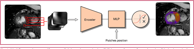
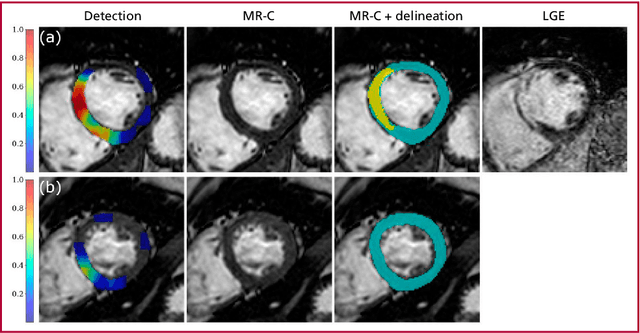
Background: Artificial intelligence techniques have demonstrated great potential in cardiology, especially to detect imperceptible patterns for the human eye. In this sense, these techniques seem to be adequate to identify patterns in the myocardial texture which could lead to characterize and quantify fibrosis. Purpose: The aim of this study was to postulate a new artificial intelligence method to identify fibrosis in cine cardiac magnetic resonance (CMR) imaging. Methods: A retrospective observational study was carried out in a population of 75 subjects from a clinical center of San Carlos de Bariloche. The proposed method analyzes the myocardial texture in cine CMR images using a convolutional neural network to determine local myocardial tissue damage. Results: An accuracy of 89% for quantifying local tissue damage was observed for the validation data set and 70% for the test set. In addition, the qualitative analysis showed a high spatial correlation in lesion location. Conclusions: The postulated method enables to spatially identify fibrosis using only the information from cine nuclear magnetic resonance studies, demonstrating the potential of this technique to quantify myocardial viability in the future or to study the lesions etiology