 Add to Chrome
 Add to Firefox
 Add to Edge
DualDiT: A Conditional Dual-Output Diffusion Transformer for Joint OCT Image and Segmentation Mask Generation
Jul 31, 2026
Background and Objective: Generating realistic medical images with anatomically accurate segmentation masks helps address the shortage of annotated data in medical imaging, particularly in optical coherence tomography (OCT) of mouse eyes, where manual retinal layer delineation is labour-intensive due to tiny structures and required expertise, resulting in scarce datasets. While diffusion models perform well in medical image synthesis, joint image-mask generation has relied mainly on U-Net-based denoisers, leaving diffusion transformers largely unexplored. Methods: We propose a conditional dual-output Diffusion Transformer (DualDiT) for joint synthesis of OCT B-scans and segmentation masks of the upper retinal cell layers in ex vivo mouse retina. DualDiT encodes both modalities into a shared latent space via a pretrained VAE, concatenates their latent representations, and performs conditional diffusion over the joint tensor. We compared DualDiT against two adapted diffusion baselines: DDPM and LDM. Generative quality was assessed via Fréchet Inception Distance (FID) and spatial FID (sFID); practical utility via synthetic data augmentation for downstream U-Net segmentation; and perceptual realism via evaluation by three domain experts. Results: DualDiT achieved the best generative quality (FID 56.14, sFID 114.35), outperforming DDPM and LDM. Expert panels misclassified 46% of synthetic samples as real and 42% of real samples as synthetic. Adding DualDiT-generated images and masks improved Dice and IoU scores on a held-out segmentation test set. Conclusions: DualDiT shows that transformer-based diffusion models can effectively learn the joint distribution of OCT images and segmentation masks, surpassing DDPM- and LDM-based baselines in generative fidelity, downstream utility, and perceptual realism, highlighting its potential for data augmentation in annotation-scarce medical imaging.

Defect Segmentation in OCT scans of ceramic parts for non-destructive inspection using deep learning
Oct 01, 2025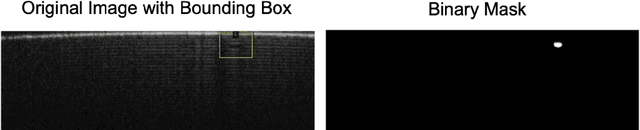
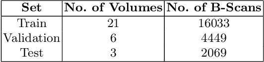
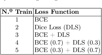
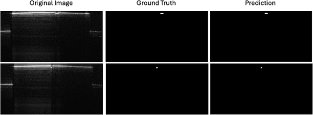
Non-destructive testing (NDT) is essential in ceramic manufacturing to ensure the quality of components without compromising their integrity. In this context, Optical Coherence Tomography (OCT) enables high-resolution internal imaging, revealing defects such as pores, delaminations, or inclusions. This paper presents an automatic defect detection system based on Deep Learning (DL), trained on OCT images with manually segmented annotations. A neural network based on the U-Net architecture is developed, evaluating multiple experimental configurations to enhance its performance. Post-processing techniques enable both quantitative and qualitative evaluation of the predictions. The system shows an accurate behavior of 0.979 Dice Score, outperforming comparable studies. The inference time of 18.98 seconds per volume supports its viability for detecting inclusions, enabling more efficient, reliable, and automated quality control.
Development of an algorithm for medical image segmentation of bone tissue in interaction with metallic implants
Apr 22, 2022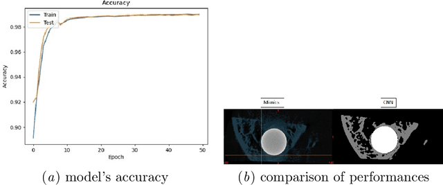
This preliminary study focuses on the development of a medical image segmentation algorithm based on artificial intelligence for calculating bone growth in contact with metallic implants. %as a result of the problem of estimating the growth of new bone tissue due to artifacts. %the presence of various types of distortions and errors, known as artifacts. Two databases consisting of computerized microtomography images have been used throughout this work: 100 images for training and 196 images for testing. Both bone and implant tissue were manually segmented in the training data set. The type of network constructed follows the U-Net architecture, a convolutional neural network explicitly used for medical image segmentation. In terms of network accuracy, the model reached around 98\%. Once the prediction was obtained from the new data set (test set), the total number of pixels belonging to bone tissue was calculated. This volume is around 15\% of the volume estimated by conventional techniques, which are usually overestimated. This method has shown its good performance and results, although it has a wide margin for improvement, modifying various parameters of the networks or using larger databases to improve training.