 Add to Chrome
 Add to Firefox
 Add to Edge
Residual Transformer Fusion Network for Salt and Pepper Image Denoising
Feb 13, 2025


Convolutional Neural Network (CNN) has been widely used in unstructured datasets, one of which is image denoising. Image denoising is a noisy image reconstruction process that aims to reduce additional noise that occurs from the noisy image with various strategies. Image denoising has a problem, namely that some image denoising methods require some prior knowledge of information about noise. To overcome this problem, a combined architecture of Convolutional Vision Transformer (CvT) and Residual Networks (ResNet) is used which is called the Residual Transformer Fusion Network (RTF-Net). In general, the process in this architecture can be divided into two parts, Noise Suppression Network (NSN) and Structure Enhancement Network (SEN). Residual Block is used in the Noise Suppression Network and is used to learn the noise map in the image, while the CvT is used in the Structure Enhancement Network and is used to learn the details that need to be added to the image processed by the Noise Suppression Network. The model was trained using the DIV2K Training Set dataset, and validation using the DIV2K Validation Set. After doing the training, the model was tested using Lena, Bridge, Pepper, and BSD300 images with noise levels ranging from 30%, 50%, and 70% and the PSNR results were compared with the DBA, NASNLM, PARIGI, NLSF, NLSF-MLP and NLSF-CNN methods. The test results show that the proposed method is superior in all cases except for Pepper's image with a noise level of 30%, where NLSF-CNN is superior with a PSNR value of 32.99 dB, while the proposed method gets a PSNR value of 31.70 dB.
Identification and Counting White Blood Cells and Red Blood Cells using Image Processing Case Study of Leukemia
Nov 16, 2015
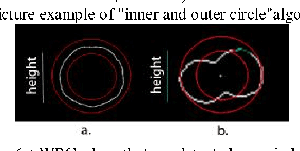
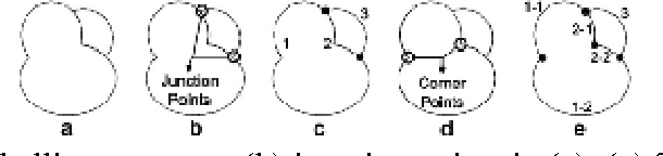
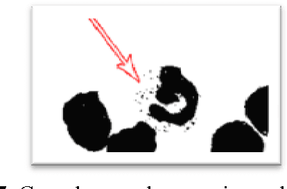
Leukemia is diagnosed with complete blood counts which is by calculating all blood cells and compare the number of white blood cells (White Blood Cells / WBC) and red blood cells (Red Blood Cells / RBC). Information obtained from a complete blood count, has become a cornerstone in the hematology laboratory for diagnostic purposes and monitoring of hematological disorders. However, the traditional procedure for counting blood cells manually requires effort and a long time, therefore this method is one of the most expensive routine tests in laboratory hematology clinic. Solution for such kind of time consuming task and necessity of data tracability can be found in image processing techniques based on blood cell morphology . This study aims to identify Acute Lymphocytic Leukemia (ALL) and Acute Myeloid Leukemia type M3 (AML M3) using Fuzzy Rule Based System based on morphology of white blood cells. Characteristic parameters witch extractedare WBC Area, Nucleus and Granule Ratio of white blood cells. Image processing algorithms such as thresholding, Canny edge detection and color identification filters are used.Then for identification of ALL, AML M3 and Healthy cells used Fuzzy Rule Based System with Sugeno method. In the testing process used 104 images out of which 29 ALL - Positive, 50 AML M3 - Positive and 25 Healthy cells. Test results showed 83.65 % accuracy .