 Add to Chrome
 Add to Firefox
 Add to Edge
Weakly supervised multiple instance learning histopathological tumor segmentation
Apr 21, 2020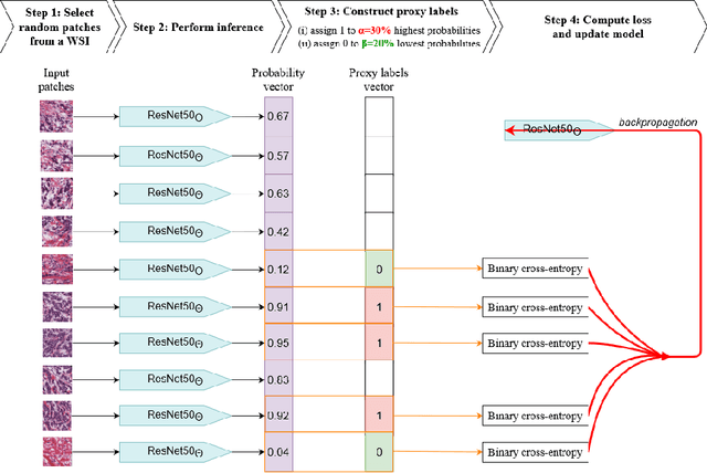
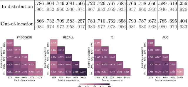
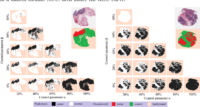
Histopathological image segmentation is a challenging and important topic in medical imaging with tremendous potential impact in clinical practice. State of the art methods relying on hand-crafted annotations that reduce the scope of the solutions since digital histology suffers from standardization and samples differ significantly between cancer phenotypes. To this end, in this paper, we propose a weakly supervised framework relying on weak standard clinical practice annotations, available in most medical centers. In particular, we exploit a multiple instance learning scheme providing a label for each instance, establishing a detailed segmentation of whole slide images. The potential of the framework is assessed with multi-centric data experiments using The Cancer Genome Atlas repository and the publicly available PatchCamelyon dataset. Promising results when compared with experts' annotations demonstrate the potentials of our approach.
AI-Driven CT-based quantification, staging and short-term outcome prediction of COVID-19 pneumonia
Apr 20, 2020Chest computed tomography (CT) is widely used for the management of Coronavirus disease 2019 (COVID-19) pneumonia because of its availability and rapidity. The standard of reference for confirming COVID-19 relies on microbiological tests but these tests might not be available in an emergency setting and their results are not immediately available, contrary to CT. In addition to its role for early diagnosis, CT has a prognostic role by allowing visually evaluating the extent of COVID-19 lung abnormalities. The objective of this study is to address prediction of short-term outcomes, especially need for mechanical ventilation. In this multi-centric study, we propose an end-to-end artificial intelligence solution for automatic quantification and prognosis assessment by combining automatic CT delineation of lung disease meeting performance of experts and data-driven identification of biomarkers for its prognosis. AI-driven combination of variables with CT-based biomarkers offers perspectives for optimal patient management given the shortage of intensive care beds and ventilators.