We present a novel multi-stage 3D computer-aided detection and diagnosis (CAD) model for automated localization of clinically significant prostate cancer (csPCa) in bi-parametric MR imaging (bpMRI). State-of-the-art attention mechanisms drive its detection network, which aims to accurately discriminate csPCa lesions from indolent cancer and the wide range of benign pathology that can afflict the prostate gland. In parallel, a decoupled residual classifier is used to achieve consistent false positive reduction, without sacrificing high detection sensitivity or computational efficiency. Furthermore, a probabilistic anatomical prior, which captures the spatial prevalence of csPCa and its zonal distinction, is computed and encoded into the CNN architecture to guide model generalization with domain-specific clinical knowledge. For 486 institutional testing scans, the 3D CAD system achieves $83.69\pm5.22\%$ and $93.19\pm2.96\%$ detection sensitivity at 0.50 and 1.46 false positive(s) per patient, respectively, along with $0.882$ AUROC in patient-based diagnosis $-$significantly outperforming four baseline architectures (USEResNet, UNet++, nnU-Net, Attention U-Net). For 296 external testing scans, the ensembled CAD system shares moderate agreement with a consensus of expert radiologists ($76.69\%$; $kappa=0.511$) and independent pathologists ($81.08\%$; $kappa=0.559$); demonstrating a strong ability to localize histologically-confirmed malignancies and generalize beyond the radiologically-estimated annotations of the 1950 training-validation cases used in this study.
We hypothesize that anatomical priors can be viable mediums to infuse domain-specific clinical knowledge into state-of-the-art convolutional neural networks (CNN) based on the U-Net architecture. We introduce a probabilistic population prior which captures the spatial prevalence and zonal distinction of clinically significant prostate cancer (csPCa), in order to improve its computer-aided detection (CAD) in bi-parametric MR imaging (bpMRI). To evaluate performance, we train 3D adaptations of the U-Net, U-SEResNet, UNet++ and Attention U-Net using 800 institutional training-validation scans, paired with radiologically-estimated annotations and our computed prior. For 200 independent testing bpMRI scans with histologically-confirmed delineations of csPCa, our proposed method of encoding clinical priori demonstrates a strong ability to improve patient-based diagnosis (upto 8.70% increase in AUROC) and lesion-level detection (average increase of 1.08 pAUC between 0.1-1.0 false positive per patient) across all four architectures.
Weakly supervised disease classification of CT imaging suffers from poor localization owing to case-level annotations, where even a positive scan can hold hundreds to thousands of negative slices along multiple planes. Furthermore, although deep learning segmentation and classification models extract distinctly unique combinations of anatomical features from the same target class(es), they are typically seen as two independent processes in a computer-aided diagnosis (CAD) pipeline, with little to no feature reuse. In this research, we propose a medical classifier that leverages the semantic structural concepts learned via multi-resolution segmentation feature maps, to guide weakly supervised 3D classification of chest CT volumes. Additionally, a comparative analysis is drawn across two different types of feature aggregation to explore the vast possibilities surrounding feature fusion. Using a dataset of 1593 scans labeled on a case-level basis via rule-based model, we train a dual-stage convolutional neural network (CNN) to perform organ segmentation and binary classification of four representative diseases (emphysema, pneumonia/atelectasis, mass and nodules) in lungs. The baseline model, with separate stages for segmentation and classification, results in AUC of 0.791. Using identical hyperparameters, the connected architecture using static and dynamic feature aggregation improves performance to AUC of 0.832 and 0.851, respectively. This study advances the field in two key ways. First, case-level report data is used to weakly supervise a 3D CT classifier of multiple, simultaneous diseases for an organ. Second, segmentation and classification models are connected with two different feature aggregation strategies to enhance the classification performance.
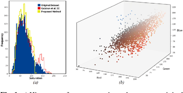
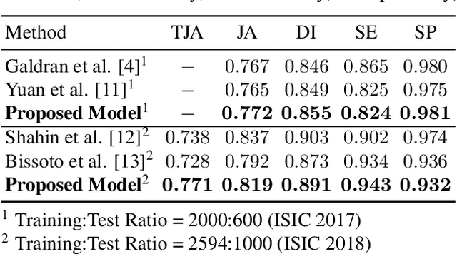
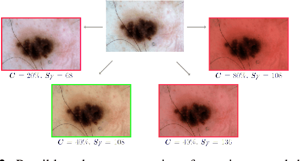
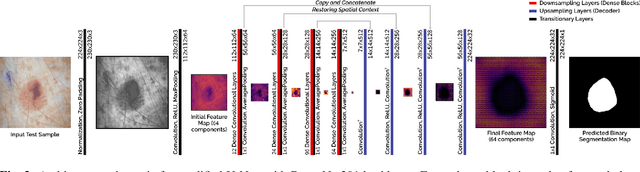
Fully automatic detection of skin lesions in dermatoscopic images can facilitate early diagnosis and repression of malignant melanoma and non-melanoma skin cancer. Although convolutional neural networks are a powerful solution, they are limited by the illumination spectrum of annotated dermatoscopic screening images, where color is an important discriminative feature. In this paper, we propose an adaptive color augmentation technique to amplify data expression and model performance, while regulating color difference and saturation to minimize the risks of using synthetic data. Through deep visualization, we qualitatively identify and verify the semantic structural features learned by the network for discriminating skin lesions against normal skin tissue. The overall system achieves a Dice Ratio of 0.891 with 0.943 sensitivity and 0.932 specificity on the ISIC 2018 Testing Set for segmentation.
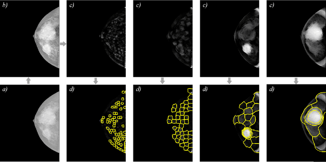
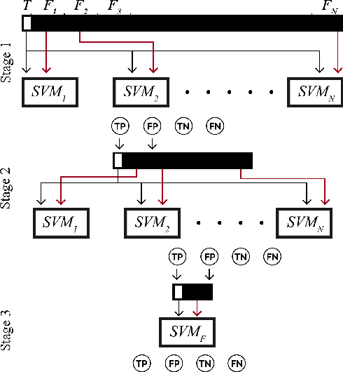
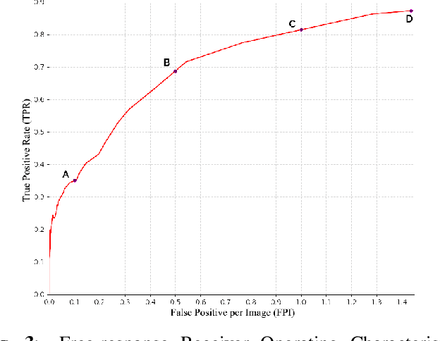
Identification and segmentation of breast masses in mammograms face complex challenges, owing to the highly variable nature of malignant densities with regards to their shape, contours, texture and orientation. Additionally, classifiers typically suffer from high class imbalance in region candidates, where normal tissue regions vastly outnumber malignant masses. This paper proposes a rigorous segmentation method, supported by morphological enhancement using grayscale linear filters. A novel cascaded ensemble of support vector machines (SVM) is used to effectively tackle the class imbalance and provide significant predictions. For True Positive Rate (TPR) of 0.35, 0.69 and 0.82, the system generates only 0.1, 0.5 and 1.0 False Positives/Image (FPI), respectively.