 Add to Chrome
 Add to Firefox
 Add to Edge
Technically Plausible but Clinically Misleading? Expert Evaluation of Patient-Personalized Synthetic Prostate MRI
Aug 17, 2026
Magnetic resonance imaging (MRI) is central to prostate cancer assessment, yet its acquisition is costly and time-consuming, making it a major bottleneck in the patient care pathway. Personalized MRI synthesis is a promising direction because it may allow the generation of clinically realistic images tailored to individual patients while reducing the dependence on scanner-acquired imaging. In this work, we in-vestigate whether patient-personalized MRI synthesis produces images that experts perceive as clinically plausible and how such personaliza-tion influences expert interpretation. We synthesize patient-personalized prostate MRI using a 3D diffusion model conditioned on routine pre-imaging clinical variables age and PSA. Using a large publicly available dataset, we first verify that synthesized images are technically compara-ble to scanner-acquired MRIs using standard image similarity metrics. We then conduct a pilot, single-blinded, randomized expert study on a subset of cases sampled to reflect variation in patient clinical pro-files. A radiologist compared personalized and unconditioned synthetic MRIs using paired assessments and Likert-scale ratings of clinical plausibility, confidence, and risk of misleading interpretation, supplemented by qualitative feedback. Although personalized synthetic MRIs appeared technically plausible, expert interpretation highlighted variability in con-fidence and potential risks of misleading cues when routine clinical data was embedded into image generation. These findings suggest that while personalized synthesis may be technically feasible, careful assessment is needed to understand how generated cues influence clinician interpreta-tion before such systems can be safely integrated into clinical workflows.

Clinical Pathways Matter for Multimodal Deep Learning in Early Alzheimers Disease Detection
Aug 17, 2026Identifying individuals at risk of Alzheimer's disease (AD), particularly in the preclinical and early stages, remains challenging. Although deep learning approaches based on structural MRI show promise as a non-invasive biomarker, existing multimodal models require task-specific training and depend on biomarkers that are not routinely available in clinical practice. Here, we propose a zero-shot multimodal framework based on SigLIP that combines structural MRI embeddings with text embeddings of routinely collected clinical variables for early AD risk stratification in individuals at preclinical or mild cognitive impairment (MCI) stages. We evaluated the approach in 416 individuals from the ADNI cohort (age: 72.73 +- 6.7). SigLIP was used without fine-tuning to extract MRI and clinical text embeddings, which were combined into multimodal representations for individual-level AD risk prediction within 4 years. We further compared the model performance in a single-visit and two-visit settings to assess the value of longitudinal information and framework scalability. In the single-visit setting, combining MRI embeddings with MMSE, age, and sex achieved an AUC of 0.91 +- 0.02, outperforming both a CSF A\b{eta}42-based model (AUC 0.73 +- 0.08) and an MMSE-based model (AUC 0.85 +- 0.22). In the two-visit setting, performance was maintained or improved, supporting the scalability of the approach to longitudinal data. These findings suggest that zero-shot multimodal fusion of structural MRI and routinely collected clinical variables may provide a practical and scalable strategy for early AD risk stratification without task-specific retraining.
Neuropsychiatric Deviations From Normative Profiles: An MRI-Derived Marker for Early Alzheimer's Disease Detection
Apr 01, 2026Neuropsychiatric symptoms (NPS) such as depression and apathy are common in Alzheimer's disease (AD) and often precede cognitive decline. NPS assessments hold promise as early detection markers due to their correlation with disease progression and their non-invasive nature. Yet current tools cannot distinguish whether NPS are part of aging or early signs of AD, limiting their utility. We present a deep learning-based normative modelling framework to identify atypical NPS burden from structural MRI. A 3D convolutional neural network was trained on cognitively stable participants from the Alzheimer's Disease Neuroimaging Initiative, learning the mapping between brain anatomy and Neuropsychiatric Inventory Questionnaire (NPIQ) scores. Deviations between predicted and observed scores defined the Divergence from NPIQ scores (DNPI). Higher DNPI was associated with future AD conversion (adjusted OR=2.5; p < 0.01) and achieved predictive accuracy comparable to cerebrospinal fluid AB42 (AUC=0.74 vs 0.75). Our approach supports scalable, non-invasive strategies for early AD detection.
Beyond the Signal: Medication State Effect on EEG-Based AI models for Parkinson's Disease
Mar 27, 2025Parkinson's disease (PD) poses a growing challenge due to its increasing prevalence, complex pathology, and functional ramifications. Electroencephalography (EEG), when integrated with artificial intelligence (AI), holds promise for monitoring disease progression, identifying sub-phenotypes, and personalizing treatment strategies. However, the effect of medication state on AI model learning and generalization remains poorly understood, potentially limiting EEG-based AI models clinical applicability. This study evaluates how medication state influences the training and generalization of EEG-based AI models. Paired EEG recordings were utilized from individuals with PD in both ON- and OFF-medication states. AI models were trained on recordings from each state separately and evaluated on independent test sets representing both ON- and OFF-medication conditions. Model performance was assessed using multiple metrics, with accuracy (ACC) as the primary outcome. Statistical significance was assessed via permutation testing (p-values<0.05). Our results reveal that models trained on OFF-medication data exhibited consistent but suboptimal performance across both medication states (ACC_OFF-ON=55.3\pm8.8 and ACC_OFF-OFF=56.2\pm8.7). In contrast, models trained on ON-medication data demonstrated significantly higher performance on ON-medication recordings (ACC_ON-ON=80.7\pm7.1) but significantly reduced generalization to OFF-medication data (ACC_ON-OFF=76.0\pm7.2). Notably, models trained on ON-medication data consistently outperformed those trained on OFF-medication data within their respective states (ACC_ON-ON=80.7\pm7.1 and ACC_OFF-OFF=56.2\pm8.7). Our findings suggest that medication state significantly influences the patterns learned by AI models. Addressing this challenge is essential to enhance the robustness and clinical utility of AI models for PD characterization and management.
Reconsidering evaluation practices in modular systems: On the propagation of errors in MRI prostate cancer detection
Sep 15, 2023Magnetic resonance imaging has evolved as a key component for prostate cancer (PCa) detection, substantially increasing the radiologist workload. Artificial intelligence (AI) systems can support radiological assessment by segmenting and classifying lesions in clinically significant (csPCa) and non-clinically significant (ncsPCa). Commonly, AI systems for PCa detection involve an automatic prostate segmentation followed by the lesion detection using the extracted prostate. However, evaluation reports are typically presented in terms of detection under the assumption of the availability of a highly accurate segmentation and an idealistic scenario, omitting the propagation of errors between modules. For that purpose, we evaluate the effect of two different segmentation networks (s1 and s2) with heterogeneous performances in the detection stage and compare it with an idealistic setting (s1:89.90+-2.23 vs 88.97+-3.06 ncsPCa, P<.001, 89.30+-4.07 and 88.12+-2.71 csPCa, P<.001). Our results depict the relevance of a holistic evaluation, accounting for all the sub-modules involved in the system.
Leveraging multi-view data without annotations for prostate MRI segmentation: A contrastive approach
Aug 12, 2023
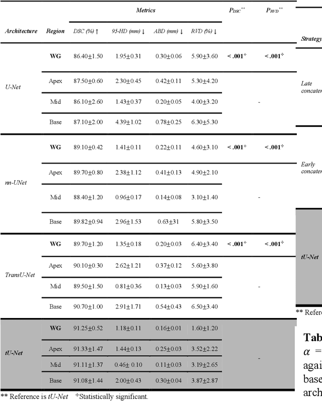
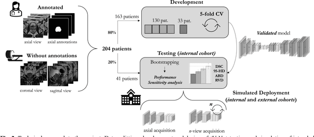
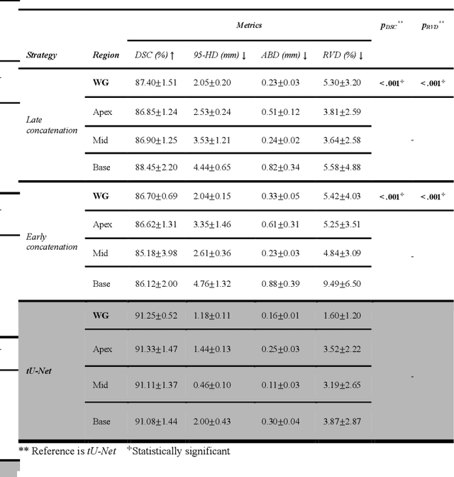
An accurate prostate delineation and volume characterization can support the clinical assessment of prostate cancer. A large amount of automatic prostate segmentation tools consider exclusively the axial MRI direction in spite of the availability as per acquisition protocols of multi-view data. Further, when multi-view data is exploited, manual annotations and availability at test time for all the views is commonly assumed. In this work, we explore a contrastive approach at training time to leverage multi-view data without annotations and provide flexibility at deployment time in the event of missing views. We propose a triplet encoder and single decoder network based on U-Net, tU-Net (triplet U-Net). Our proposed architecture is able to exploit non-annotated sagittal and coronal views via contrastive learning to improve the segmentation from a volumetric perspective. For that purpose, we introduce the concept of inter-view similarity in the latent space. To guide the training, we combine a dice score loss calculated with respect to the axial view and its manual annotations together with a multi-view contrastive loss. tU-Net shows statistical improvement in dice score coefficient (DSC) with respect to only axial view (91.25+-0.52% compared to 86.40+-1.50%,P<.001). Sensitivity analysis reveals the volumetric positive impact of the contrastive loss when paired with tU-Net (2.85+-1.34% compared to 3.81+-1.88%,P<.001). Further, our approach shows good external volumetric generalization in an in-house dataset when tested with multi-view data (2.76+-1.89% compared to 3.92+-3.31%,P=.002), showing the feasibility of exploiting non-annotated multi-view data through contrastive learning whilst providing flexibility at deployment in the event of missing views.
Out-of-distribution multi-view auto-encoders for prostate cancer lesion detection
Aug 12, 2023

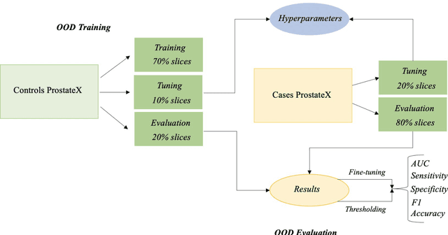
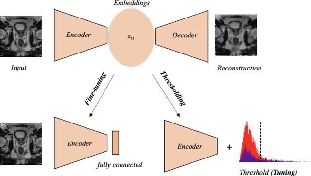
Traditional deep learning (DL) approaches based on supervised learning paradigms require large amounts of annotated data that are rarely available in the medical domain. Unsupervised Out-of-distribution (OOD) detection is an alternative that requires less annotated data. Further, OOD applications exploit the class skewness commonly present in medical data. Magnetic resonance imaging (MRI) has proven to be useful for prostate cancer (PCa) diagnosis and management, but current DL approaches rely on T2w axial MRI, which suffers from low out-of-plane resolution. We propose a multi-stream approach to accommodate different T2w directions to improve the performance of PCa lesion detection in an OOD approach. We evaluate our approach on a publicly available data-set, obtaining better detection results in terms of AUC when compared to a single direction approach (73.1 vs 82.3). Our results show the potential of OOD approaches for PCa lesion detection based on MRI.
Prostate Age Gap : An MRI surrogate marker of aging for prostate cancer detection
Aug 10, 2023Background: Prostate cancer (PC) MRI-based risk calculators are commonly based on biological (e.g. PSA), MRI markers (e.g. volume), and patient age. Whilst patient age measures the amount of years an individual has existed, biological age (BA) might better reflect the physiology of an individual. However, surrogates from prostate MRI and linkage with clinically significant PC (csPC) remain to be explored. Purpose: To obtain and evaluate Prostate Age Gap (PAG) as an MRI marker tool for csPC risk. Study type: Retrospective. Population: A total of 7243 prostate MRI slices from 468 participants who had undergone prostate biopsies. A deep learning model was trained on 3223 MRI slices cropped around the gland from 81 low-grade PC (ncsPC, Gleason score <=6) and 131 negative cases and tested on the remaining 256 participants. Assessment: Chronological age was defined as the age of the participant at the time of the visit and used to train the deep learning model to predict the age of the patient. Following, we obtained PAG, defined as the model predicted age minus the patient's chronological age. Multivariate logistic regression models were used to estimate the association through odds ratio (OR) and predictive value of PAG and compared against PSA levels and PI-RADS>=3. Statistical tests: T-test, Mann-Whitney U test, Permutation test and ROC curve analysis. Results: The multivariate adjusted model showed a significant difference in the odds of clinically significant PC (csPC, Gleason score >=7) (OR =3.78, 95% confidence interval (CI):2.32-6.16, P <.001). PAG showed a better predictive ability when compared to PI-RADS>=3 and adjusted by other risk factors, including PSA levels: AUC =0.981 vs AUC =0.704, p<.001. Conclusion: PAG was significantly associated with the risk of clinically significant PC and outperformed other well-established PC risk factors.
Assessing gender fairness in EEG-based machine learning detection of Parkinson's disease: A multi-center study
Mar 11, 2023


As the number of automatic tools based on machine learning (ML) and resting-state electroencephalography (rs-EEG) for Parkinson's disease (PD) detection keeps growing, the assessment of possible exacerbation of health disparities by means of fairness and bias analysis becomes more relevant. Protected attributes, such as gender, play an important role in PD diagnosis development. However, analysis of sub-group populations stemming from different genders is seldom taken into consideration in ML models' development or the performance assessment for PD detection. In this work, we perform a systematic analysis of the detection ability for gender sub-groups in a multi-center setting of a previously developed ML algorithm based on power spectral density (PSD) features of rs-EEG. We find significant differences in the PD detection ability for males and females at testing time (80.5% vs. 63.7% accuracy) and significantly higher activity for a set of parietal and frontal EEG channels and frequency sub-bands for PD and non-PD males that might explain the differences in the PD detection ability for the gender sub-groups.
Machine Learning-Based Detection of Parkinson's Disease From Resting-State EEG: A Multi-Center Study
Mar 02, 2023Resting-state EEG (rs-EEG) has been demonstrated to aid in Parkinson's disease (PD) diagnosis. In particular, the power spectral density (PSD) of low-frequency bands ({\delta} and {\theta}) and high-frequency bands ({\alpha} and \b{eta}) has been shown to be significantly different in patients with PD as compared to subjects without PD (non-PD). However, rs-EEG feature extraction and the interpretation thereof can be time-intensive and prone to examiner variability. Machine learning (ML) has the potential to automatize the analysis of rs-EEG recordings and provides a supportive tool for clinicians to ease their workload. In this work, we use rs-EEG recordings of 84 PD and 85 non-PD subjects pooled from four datasets obtained at different centers. We propose an end-to-end pipeline consisting of preprocessing, extraction of PSD features from clinically validated frequency bands, and feature selection before evaluating the classification ability of the features via ML algorithms to stratify between PD and non-PD subjects. Further, we evaluate the effect of feature harmonization, given the multi-center nature of the datasets. Our validation results show, on average, an improvement in PD detection ability (69.6% vs. 75.5% accuracy) by logistic regression when harmonizing the features and performing univariate feature selection (k = 202 features). Our final results show an average global accuracy of 72.2% with balanced accuracy results for all the centers included in the study: 60.6%, 68.7%, 77.7%, and 82.2%, respectively.