 Add to Chrome
 Add to Firefox
 Add to Edge
Automated ACL Footprint Identification Using 3D Deep Learning
Aug 18, 2026
One of the most common reasons for anterior cruciate ligament (ACL) reconstruction failure is femoral tunnel malpositioning (ACL footprint center and tunnel orientation). Such failures may lead to the development of meniscal pathology and osteoarthritis. Accurate ACL femoral footprint identification is therefore essential for precise tunnel placement, restoration of the native knee joint mechanics, post-surgical knee joint health and prevention of graft failure. Recent advances in artificial intelligence (AI) bring new opportunities to improve image-guided orthopedic surgery. However, at present, existing AI research focuses primarily on ACL segmentation and rupture classification based on pre- and post-operative magnetic resonance (MR) images. Identification of the ACL footprint center using deep learning methods has not been thoroughly researched. Thus, the purpose of this study is to explore 3D deep learning models for ACL femoral footprint identification directly from 3D MR images. Two comprehensive 3D deep learning architectures were developed: a 3D graph convolutional neural network-based geometric model applied to 3D femoral meshes; and a 3D landmark-enhanced identification model based on 3D MR images. A total of 4883 right and 3087 left knee image sets were used from a publicly available database. Eighty percent (80%) were applied to model generation, and twenty percent (20%) were preserved for model testing. Both models achieved excellent performance; however, the image-based method outperformed the model-based method (average error of 2.1mm vs 2.8 mm). Thus, 3D deep learning provides a feasible clinical approach for ACL footprint localization and has the potential to improve ACL reconstruction footprint accuracy.

RTA-Former: Reverse Transformer Attention for Polyp Segmentation
Jan 22, 2024Polyp segmentation is a key aspect of colorectal cancer prevention, enabling early detection and guiding subsequent treatments. Intelligent diagnostic tools, including deep learning solutions, are widely explored to streamline and potentially automate this process. However, even with many powerful network architectures, there still comes the problem of producing accurate edge segmentation. In this paper, we introduce a novel network, namely RTA-Former, that employs a transformer model as the encoder backbone and innovatively adapts Reverse Attention (RA) with a transformer stage in the decoder for enhanced edge segmentation. The results of the experiments illustrate that RTA-Former achieves state-of-the-art (SOTA) performance in five polyp segmentation datasets. The strong capability of RTA-Former holds promise in improving the accuracy of Transformer-based polyp segmentation, potentially leading to better clinical decisions and patient outcomes. Our code will be publicly available on GitHub.
Using Uncertainty in Deep Learning Reconstruction for Cone-Beam CT of the Brain
Aug 20, 2021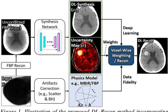
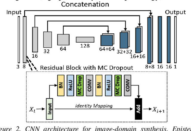
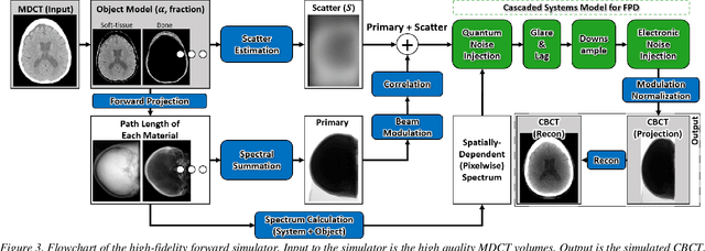
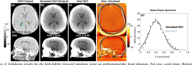
Contrast resolution beyond the limits of conventional cone-beam CT (CBCT) systems is essential to high-quality imaging of the brain. We present a deep learning reconstruction method (dubbed DL-Recon) that integrates physically principled reconstruction models with DL-based image synthesis based on the statistical uncertainty in the synthesis image. A synthesis network was developed to generate a synthesized CBCT image (DL-Synthesis) from an uncorrected filtered back-projection (FBP) image. To improve generalizability (including accurate representation of lesions not seen in training), voxel-wise epistemic uncertainty of DL-Synthesis was computed using a Bayesian inference technique (Monte-Carlo dropout). In regions of high uncertainty, the DL-Recon method incorporates information from a physics-based reconstruction model and artifact-corrected projection data. Two forms of the DL-Recon method are proposed: (i) image-domain fusion of DL-Synthesis and FBP (DL-FBP) weighted by DL uncertainty; and (ii) a model-based iterative image reconstruction (MBIR) optimization using DL-Synthesis to compute a spatially varying regularization term based on DL uncertainty (DL-MBIR). The error in DL-Synthesis images was correlated with the uncertainty in the synthesis estimate. Compared to FBP and PWLS, the DL-Recon methods (both DL-FBP and DL-MBIR) showed ~50% reduction in noise (at matched spatial resolution) and ~40-70% improvement in image uniformity. Conventional DL-Synthesis alone exhibited ~10-60% under-estimation of lesion contrast and ~5-40% reduction in lesion segmentation accuracy (Dice coefficient) in simulated and real brain lesions, suggesting a lack of reliability / generalizability for structures unseen in the training data. DL-FBP and DL-MBIR improved the accuracy of reconstruction by directly incorporating information from the measurements in regions of high uncertainty.