 Add to Chrome
 Add to Firefox
 Add to Edge
Multi-classification of High-Frequency Oscillations Using iEEG Signals and Deep Learning Models
Dec 22, 2024


Over the past decade, high-frequency oscillations (HFOs) have been studied as a promising biomarker for localizing epileptogenic areas in drug-resistant patients requiring pre-surgical intervention, while exploiting intracranial electroencephalographic iEEG. Consequently, it's important to develop accurate methods for predicting epileptic seizures. Seizure prediction involves classifying appropriate indicators, which is a difficult classification problem. Deep learning techniques, such as convolutional neural networks (CNN), have shown great promise in analyzing and classifying epilepsy-related iEEG signals. In this study, we proposed three global methods, multiclass SVM, multiple architecture CNN, and CNN-SVM, which are evaluated on a simulated iEEG dataset and then on a real iEEG signal. Our best results for the three models yield high accuracy rates, GoogLeNet-SVM achieves approximately 99.63% and 94.07% for simulated data (1) and real data (2), respectively, SVM multiclass achieves 98.14% and 88.51% for (1) and (2), respectively, and GoogLeNet achieves 98.52% and 91.85% for (1) and (2), respectively. Furthermore, the proposed model performs better than other current techniques. These results suggest that deep learning models could be a successful strategy for classifying epilepsy indicators and could potentially improve seizure prediction methods, thus improving the life quality of epileptic patients.
Comparison of inverse problem linear and non-linear methods for localization source: a combined TMS-EEG study
Nov 30, 2021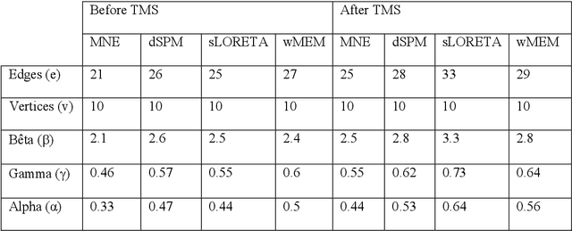
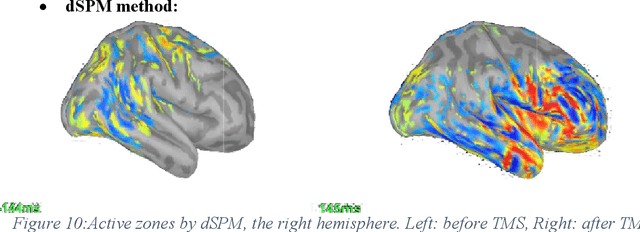
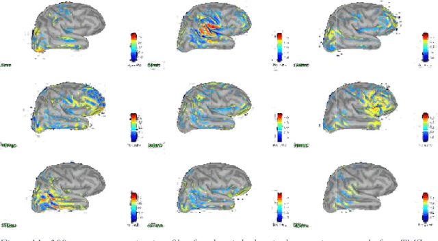
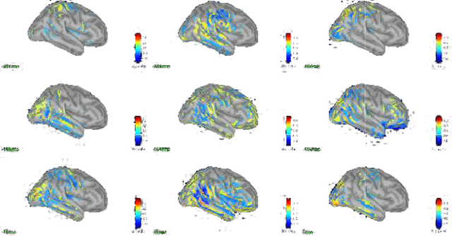
The Electro-Encephalo-Graphy (EEG) technique consists of estimating the cortical distribution of signals over time of electrical activity and also of locating the zones of primary sensory projection. Moreover, it is able to record respectively the variations of potential and field magnetic waves generated by electrical activity in the brain every millisecond. Concerning, the study of the localization source, the brain localizationactivity requires the solution of a inverse problem. Many different imaging methods are used to solve the inverse problem.The aim of the presentstudy is to provide comparison criteria for choosing the least bad method. Hence, the transcranial magnetic stimulation (TMS) and electroencephalography (EEG) technique are combined for the sake of studying the dynamics of the brain at rest following a disturbance. The study focuses in the comparison of the following methods for EEG following stimulation by TMS: sLORETA (standardized Low Resolution Electromagnetic Tomography), MNE (Minimum Estimate of the standard), dSPM (dynamic Statistical Parametric Mapping) and wMEM (wavelet based on the Maximum Entropy on the Mean)in order to study the impact of TMS towards rest and to study inter and intra zone connectivity.The contribution of the comparison is demonstrated via the stages of the simulations.