 Add to Chrome
 Add to Firefox
 Add to Edge
"cancer detection": models, code, and papers
A Comprehensive Study of Data Augmentation Strategies for Prostate Cancer Detection in Diffusion-weighted MRI using Convolutional Neural Networks
Jun 01, 2020
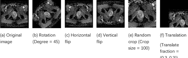
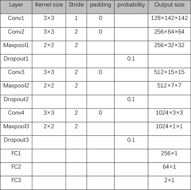
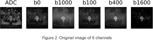
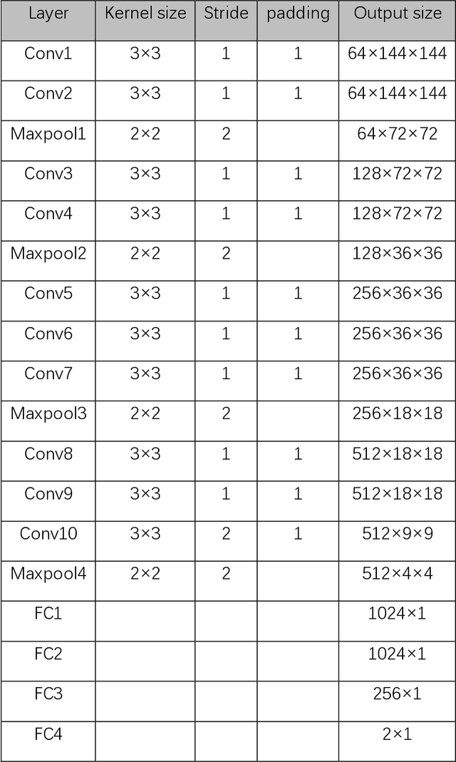
Data augmentation refers to a group of techniques whose goal is to battle limited amount of available data to improve model generalization and push sample distribution toward the true distribution. While different augmentation strategies and their combinations have been investigated for various computer vision tasks in the context of deep learning, a specific work in the domain of medical imaging is rare and to the best of our knowledge, there has been no dedicated work on exploring the effects of various augmentation methods on the performance of deep learning models in prostate cancer detection. In this work, we have statically applied five most frequently used augmentation techniques (random rotation, horizontal flip, vertical flip, random crop, and translation) to prostate Diffusion-weighted Magnetic Resonance Imaging training dataset of 217 patients separately and evaluated the effect of each method on the accuracy of prostate cancer detection. The augmentation algorithms were applied independently to each data channel and a shallow as well as a deep Convolutional Neural Network (CNN) were trained on the five augmented sets separately. We used Area Under Receiver Operating Characteristic (ROC) curve (AUC) to evaluate the performance of the trained CNNs on a separate test set of 95 patients, using a validation set of 102 patients for finetuning. The shallow network outperformed the deep network with the best 2D slice-based AUC of 0.85 obtained by the rotation method.
Context-Aware Transformers For Spinal Cancer Detection and Radiological Grading
Jun 27, 2022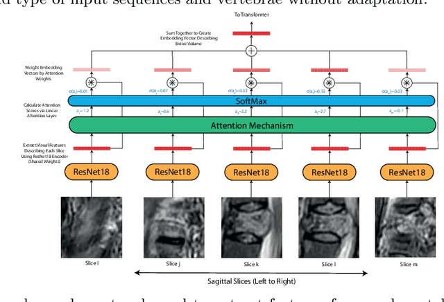
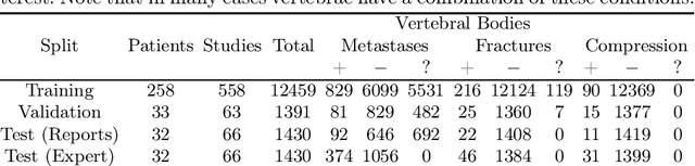
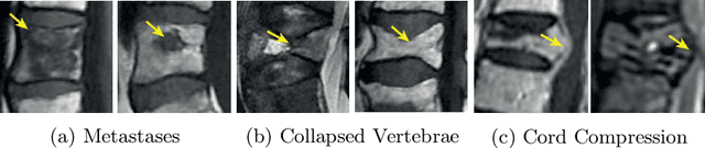
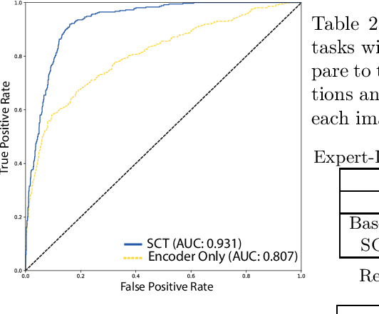
This paper proposes a novel transformer-based model architecture for medical imaging problems involving analysis of vertebrae. It considers two applications of such models in MR images: (a) detection of spinal metastases and the related conditions of vertebral fractures and metastatic cord compression, (b) radiological grading of common degenerative changes in intervertebral discs. Our contributions are as follows: (i) We propose a Spinal Context Transformer (SCT), a deep-learning architecture suited for the analysis of repeated anatomical structures in medical imaging such as vertebral bodies (VBs). Unlike previous related methods, SCT considers all VBs as viewed in all available image modalities together, making predictions for each based on context from the rest of the spinal column and all available imaging modalities. (ii) We apply the architecture to a novel and important task: detecting spinal metastases and the related conditions of cord compression and vertebral fractures/collapse from multi-series spinal MR scans. This is done using annotations extracted from free-text radiological reports as opposed to bespoke annotation. However, the resulting model shows strong agreement with vertebral-level bespoke radiologist annotations on the test set. (iii) We also apply SCT to an existing problem: radiological grading of inter-vertebral discs (IVDs) in lumbar MR scans for common degenerative changes.We show that by considering the context of vertebral bodies in the image, SCT improves the accuracy for several gradings compared to previously published model.
A Method to Facilitate Cancer Detection and Type Classification from Gene Expression Data using a Deep Autoencoder and Neural Network
Dec 20, 2018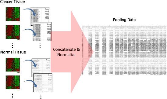
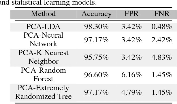
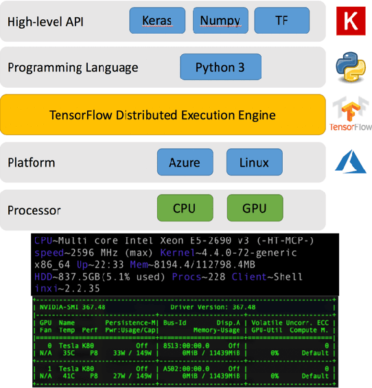
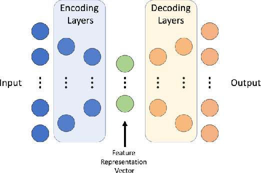
With the increased affordability and availability of whole-genome sequencing, large-scale and high-throughput gene expression is widely used to characterize diseases, including cancers. However, establishing specificity in cancer diagnosis using gene expression data continues to pose challenges due to the high dimensionality and complexity of the data. Here we present models of deep learning (DL) and apply them to gene expression data for the diagnosis and categorization of cancer. In this study, we have developed two DL models using messenger ribonucleic acid (mRNA) datasets available from the Genomic Data Commons repository. Our models achieved 98% accuracy in cancer detection, with false negative and false positive rates below 1.7%. In our results, we demonstrated that 18 out of 32 cancer-typing classifications achieved more than 90% accuracy. Due to the limitation of a small sample size (less than 50 observations), certain cancers could not achieve a higher accuracy in typing classification, but still achieved high accuracy for the cancer detection task. To validate our models, we compared them with traditional statistical models. The main advantage of our models over traditional cancer detection is the ability to use data from various cancer types to automatically form features to enhance the detection and diagnosis of a specific cancer type.
Deep Learning Hyperparameter Optimization for Breast Mass Detection in Mammograms
Jul 22, 2022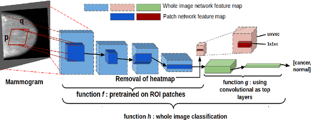
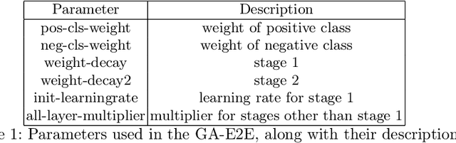
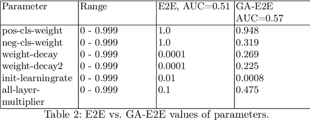
Accurate breast cancer diagnosis through mammography has the potential to save millions of lives around the world. Deep learning (DL) methods have shown to be very effective for mass detection in mammograms. Additional improvements of current DL models will further improve the effectiveness of these methods. A critical issue in this context is how to pick the right hyperparameters for DL models. In this paper, we present GA-E2E, a new approach for tuning the hyperparameters of DL models for brest cancer detection using Genetic Algorithms (GAs). Our findings reveal that differences in parameter values can considerably alter the area under the curve (AUC), which is used to determine a classifier's performance.
Enhancing Early Lung Cancer Detection on Chest Radiographs with AI-assistance: A Multi-Reader Study
Aug 31, 2022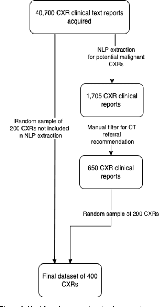
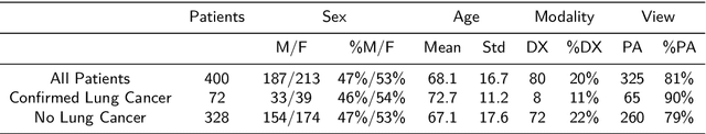
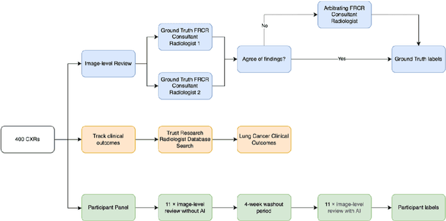
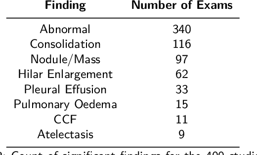
Objectives: The present study evaluated the impact of a commercially available explainable AI algorithm in augmenting the ability of clinicians to identify lung cancer on chest X-rays (CXR). Design: This retrospective study evaluated the performance of 11 clinicians for detecting lung cancer from chest radiographs, with and without assistance from a commercially available AI algorithm (red dot, Behold.ai) that predicts suspected lung cancer from CXRs. Clinician performance was evaluated against clinically confirmed diagnoses. Setting: The study analysed anonymised patient data from an NHS hospital; the dataset consisted of 400 chest radiographs from adult patients (18 years and above) who had a CXR performed in 2020, with corresponding clinical text reports. Participants: A panel of readers consisting of 11 clinicians (consultant radiologists, radiologist trainees and reporting radiographers) participated in this study. Main outcome measures: Overall accuracy, sensitivity, specificity and precision for detecting lung cancer on CXRs by clinicians, with and without AI input. Agreement rates between clinicians and performance standard deviation were also evaluated, with and without AI input. Results: The use of the AI algorithm by clinicians led to an improved overall performance for lung tumour detection, achieving an overall increase of 17.4% of lung cancers being identified on CXRs which would have otherwise been missed, an overall increase in detection of smaller tumours, a 24% and 13% increased detection of stage 1 and stage 2 lung cancers respectively, and standardisation of clinician performance. Conclusions: This study showed great promise in the clinical utility of AI algorithms in improving early lung cancer diagnosis and promoting health equity through overall improvement in reader performances, without impacting downstream imaging resources.
Genetic Analysis of Prostate Cancer with Computer Science Methods
Mar 29, 2023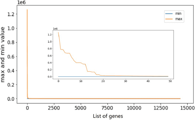
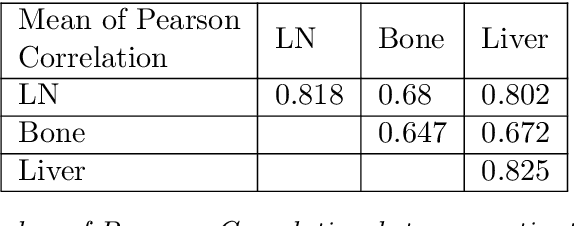
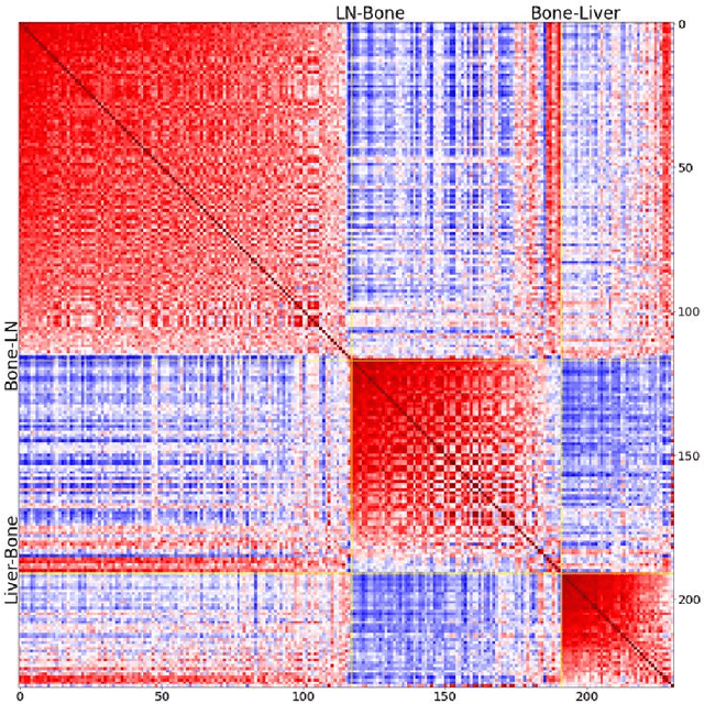
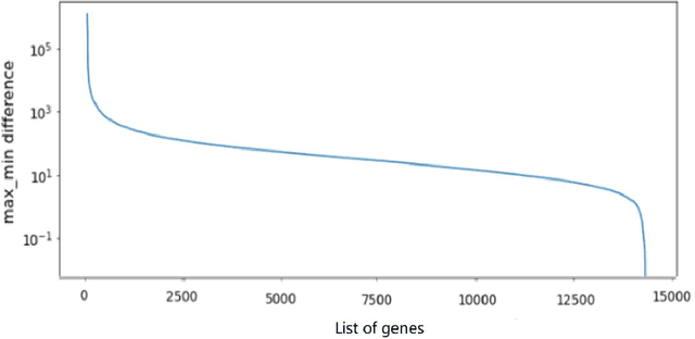
Metastatic prostate cancer is one of the most common cancers in men. In the advanced stages of prostate cancer, tumours can metastasise to other tissues in the body, which is fatal. In this thesis, we performed a genetic analysis of prostate cancer tumours at different metastatic sites using data science, machine learning and topological network analysis methods. We presented a general procedure for pre-processing gene expression datasets and pre-filtering significant genes by analytical methods. We then used machine learning models for further key gene filtering and secondary site tumour classification. Finally, we performed gene co-expression network analysis and community detection on samples from different prostate cancer secondary site types. In this work, 13 of the 14,379 genes were selected as the most metastatic prostate cancer related genes, achieving approximately 92% accuracy under cross-validation. In addition, we provide preliminary insights into the co-expression patterns of genes in gene co-expression networks. Project code is available at https://github.com/zcablii/Master_cancer_project.
Circular Antenna Array Design for Breast Cancer Detection
Jan 15, 2018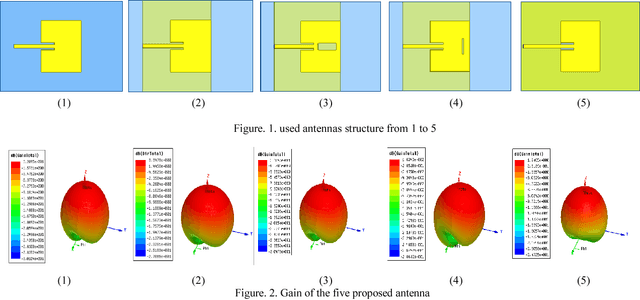
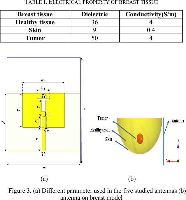
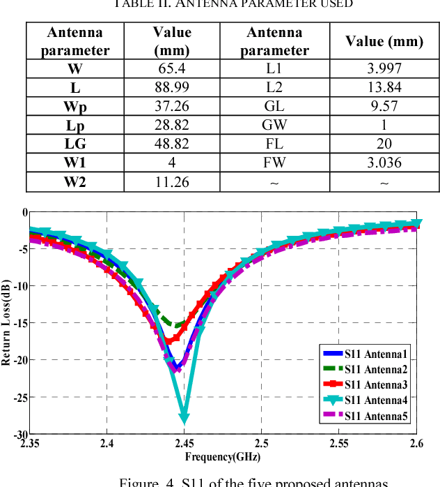
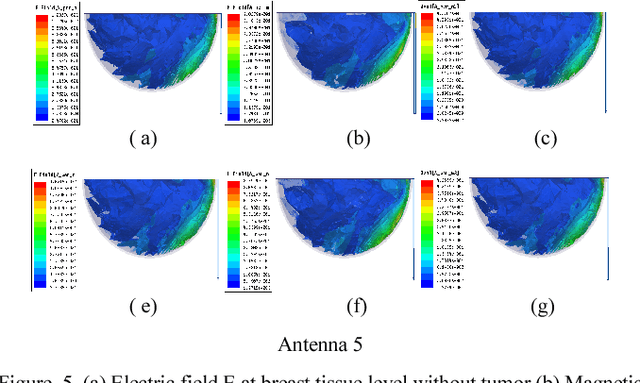
Microwave imaging for breast cancer detection is based on the contrast in the electrical properties of healthy fatty breast tissues. This paper presents an industrial, scientific and medical (ISM) bands comparative study of five microstrip patch antennas for microwave imaging at a frequency of 2.45 GHz. The choice of one antenna is made for an antenna array composed of 8 antennas for a microwave breast imaging system. Each antenna element is arranged in a circular configuration so that it can be directly faced to the breast phantom for better tumor detection. This choice is made by putting each antenna alone on the Breast skin to study the electric field, magnetic fields and current density in the healthy tissue of the breast phantom designed and simulated in Ansoft High Frequency Simulation Software (HFSS).
RADIFUSION: A multi-radiomics deep learning based breast cancer risk prediction model using sequential mammographic images with image attention and bilateral asymmetry refinement
Apr 01, 2023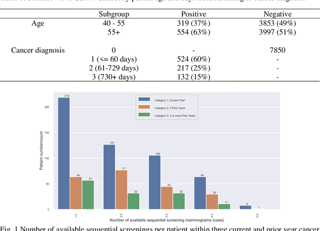
Breast cancer is a significant public health concern and early detection is critical for triaging high risk patients. Sequential screening mammograms can provide important spatiotemporal information about changes in breast tissue over time. In this study, we propose a deep learning architecture called RADIFUSION that utilizes sequential mammograms and incorporates a linear image attention mechanism, radiomic features, a new gating mechanism to combine different mammographic views, and bilateral asymmetry-based finetuning for breast cancer risk assessment. We evaluate our model on a screening dataset called Cohort of Screen-Aged Women (CSAW) dataset. Based on results obtained on the independent testing set consisting of 1,749 women, our approach achieved superior performance compared to other state-of-the-art models with area under the receiver operating characteristic curves (AUCs) of 0.905, 0.872 and 0.866 in the three respective metrics of 1-year AUC, 2-year AUC and > 2-year AUC. Our study highlights the importance of incorporating various deep learning mechanisms, such as image attention, radiomic features, gating mechanism, and bilateral asymmetry-based fine-tuning, to improve the accuracy of breast cancer risk assessment. We also demonstrate that our model's performance was enhanced by leveraging spatiotemporal information from sequential mammograms. Our findings suggest that RADIFUSION can provide clinicians with a powerful tool for breast cancer risk assessment.
EMT-NET: Efficient multitask network for computer-aided diagnosis of breast cancer
Jan 13, 2022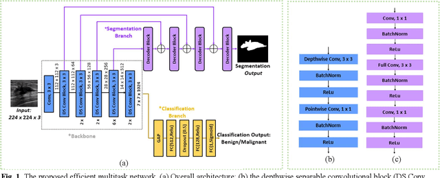
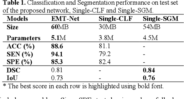
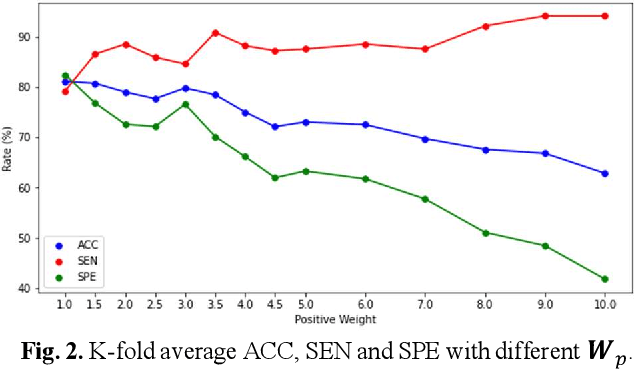
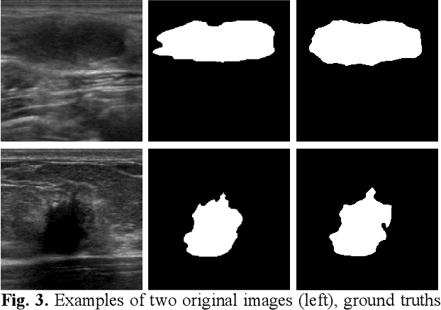
Deep learning-based computer-aided diagnosis has achieved unprecedented performance in breast cancer detection. However, most approaches are computationally intensive, which impedes their broader dissemination in real-world applications. In this work, we propose an efficient and light-weighted multitask learning architecture to classify and segment breast tumors simultaneously. We incorporate a segmentation task into a tumor classification network, which makes the backbone network learn representations focused on tumor regions. Moreover, we propose a new numerically stable loss function that easily controls the balance between the sensitivity and specificity of cancer detection. The proposed approach is evaluated using a breast ultrasound dataset with 1,511 images. The accuracy, sensitivity, and specificity of tumor classification is 88.6%, 94.1%, and 85.3%, respectively. We validate the model using a virtual mobile device, and the average inference time is 0.35 seconds per image.
Fluorescence molecular optomic signatures improve identification of tumors in head and neck specimens
Aug 29, 2022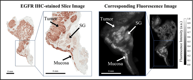
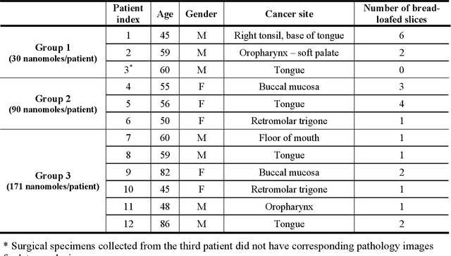
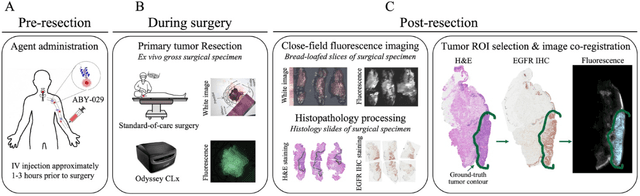
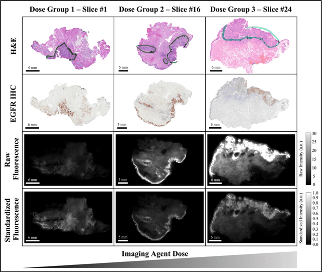
In this study, a radiomics approach was extended to optical fluorescence molecular imaging data for tissue classification, termed 'optomics'. Fluorescence molecular imaging is emerging for precise surgical guidance during head and neck squamous cell carcinoma (HNSCC) resection. However, the tumor-to-normal tissue contrast is confounded by intrinsic physiological limitations of heterogeneous expression of the target molecule, epidermal growth factor receptor (EGFR). Optomics seek to improve tumor identification by probing textural pattern differences in EGFR expression conveyed by fluorescence. A total of 1,472 standardized optomic features were extracted from fluorescence image samples. A supervised machine learning pipeline involving a support vector machine classifier was trained with 25 top-ranked features selected by minimum redundancy maximum relevance criterion. Model predictive performance was compared to fluorescence intensity thresholding method by classifying testing set image patches of resected tissue with histologically confirmed malignancy status. The optomics approach provided consistent improvement in prediction accuracy on all test set samples, irrespective of dose, compared to fluorescence intensity thresholding method (mean accuracies of 89% vs. 81%; P = 0.0072). The improved performance demonstrates that extending the radiomics approach to fluorescence molecular imaging data offers a promising image analysis technique for cancer detection in fluorescence-guided surgery.