 Add to Chrome
 Add to Firefox
 Add to Edge
"Image": models, code, and papers
Cross-Task Representation Learning for Anatomical Landmark Detection
Sep 28, 2020
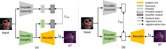
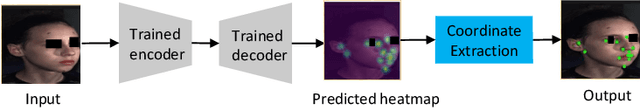
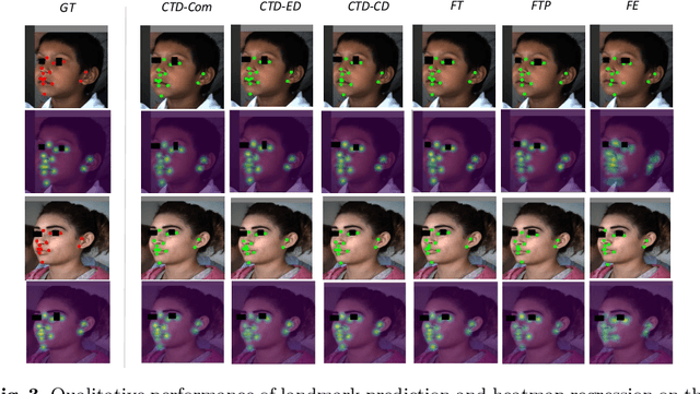
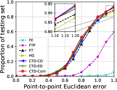
Recently, there is an increasing demand for automatically detecting anatomical landmarks which provide rich structural information to facilitate subsequent medical image analysis. Current methods related to this task often leverage the power of deep neural networks, while a major challenge in fine tuning such models in medical applications arises from insufficient number of labeled samples. To address this, we propose to regularize the knowledge transfer across source and target tasks through cross-task representation learning. The proposed method is demonstrated for extracting facial anatomical landmarks which facilitate the diagnosis of fetal alcohol syndrome. The source and target tasks in this work are face recognition and landmark detection, respectively. The main idea of the proposed method is to retain the feature representations of the source model on the target task data, and to leverage them as an additional source of supervisory signals for regularizing the target model learning, thereby improving its performance under limited training samples. Concretely, we present two approaches for the proposed representation learning by constraining either final or intermediate model features on the target model. Experimental results on a clinical face image dataset demonstrate that the proposed approach works well with few labeled data, and outperforms other compared approaches.
NeMo: Neural Mesh Models of Contrastive Features for Robust 3D Pose Estimation
Jan 29, 2021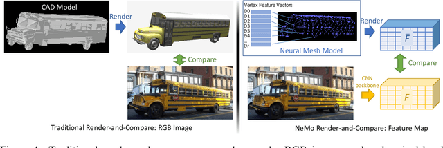
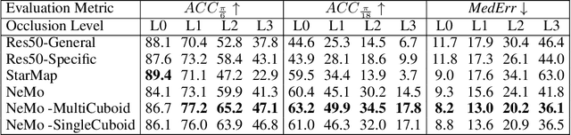
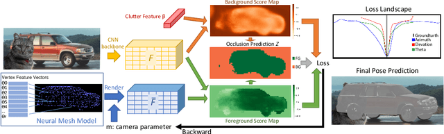
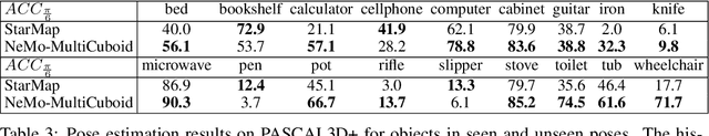
3D pose estimation is a challenging but important task in computer vision. In this work, we show that standard deep learning approaches to 3D pose estimation are not robust when objects are partially occluded or viewed from a previously unseen pose. Inspired by the robustness of generative vision models to partial occlusion, we propose to integrate deep neural networks with 3D generative representations of objects into a unified neural architecture that we term NeMo. In particular, NeMo learns a generative model of neural feature activations at each vertex on a dense 3D mesh. Using differentiable rendering we estimate the 3D object pose by minimizing the reconstruction error between NeMo and the feature representation of the target image. To avoid local optima in the reconstruction loss, we train the feature extractor to maximize the distance between the individual feature representations on the mesh using contrastive learning. Our extensive experiments on PASCAL3D+, occluded-PASCAL3D+ and ObjectNet3D show that NeMo is much more robust to partial occlusion and unseen pose compared to standard deep networks, while retaining competitive performance on regular data. Interestingly, our experiments also show that NeMo performs reasonably well even when the mesh representation only crudely approximates the true object geometry with a cuboid, hence revealing that the detailed 3D geometry is not needed for accurate 3D pose estimation. The code is publicly available at https://github.com/Angtian/NeMo.
Stabilizing Deep Tomographic Reconstruction Networks
Aug 19, 2020
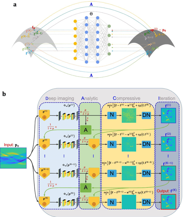
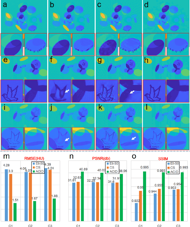
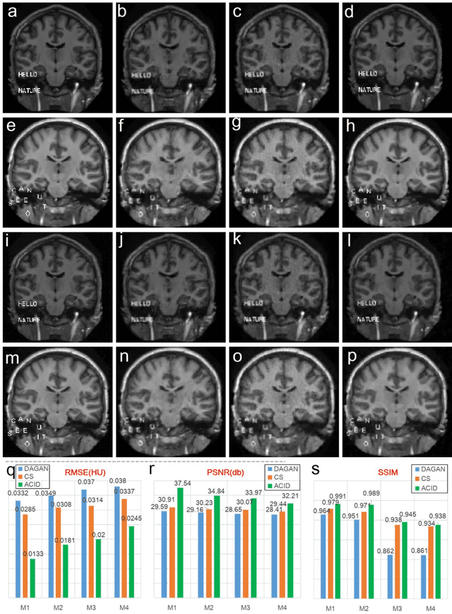
Tomographic image reconstruction with deep learning is an emerging field of applied artificial intelligence but a recent study reveals that deep reconstruction networks, such as well-known AUTOMAP, are unstable for computed tomography (CT) and magnetic resonance imaging (MRI). Specifically, three kinds of instabilities were identified: (1) strong output artefacts from tiny perturbation, (2) poor detection of small features, and (3) decreased performance with increased input data. These instabilities are believed to be from lacking kernel awareness and nontrivial to overcome, but compressed sensing (CS) reconstruction was reported to be stable due to its kernel awareness. Since deep reconstruction may potentially become the main driving force to achieve better image quality, stabilizing deep reconstruction networks is an urgent challenge. Here we propose an Analytic, Compressive, Iterative Deep (ACID) network to fundamentally address this challenge. Instead of only using deep learning or compressed sensing, ACID consists of four modules including deep reconstruction, CS, analytic mapping, and iterative refinement. In our experiments, ACID eliminated all three kinds of instabilities and significantly improved image quality relative to the methods in the aforementioned PNAS study. ACID is only an example of integrating diverse algorithmic ingredients but it has clearly demonstrated that data-driven reconstruction can be stabilized to outperform reconstruction using CS alone. The power of ACID comes from a unique combination of a deep reconstruction network trained on big data, CS via advanced optimization, and iterative refinement that stabilizes the whole workflow. We anticipate that this integrative closed-loop data driven approach will add great value to clinical and other applications.
A Systematic Approach for MRI Brain Tumor Localization, and Segmentation using Deep Learning and Active Contouring
Feb 06, 2021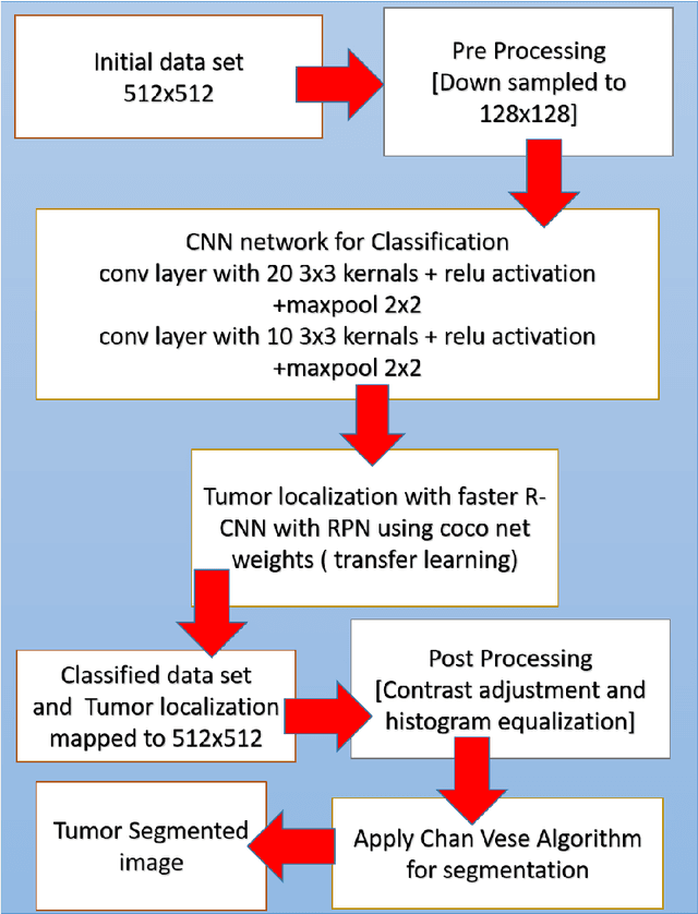
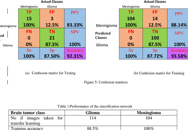
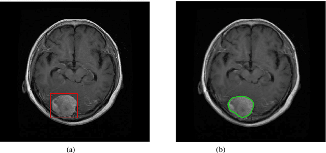
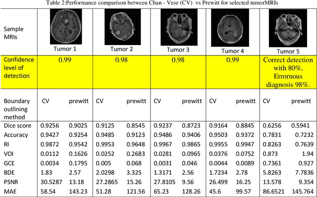
One of the main requirements of tumor extraction is the annotation and segmentation of tumor boundaries correctly. For this purpose, we present a threefold deep learning architecture. First classifiers are implemented with a deep convolutional neural network(CNN) andsecond a region-based convolutional neural network (R-CNN) is performed on the classified images to localize the tumor regions of interest. As the third and final stage, the concentratedtumor boundary is contoured for the segmentation process by using the Chan-Vesesegmentation algorithm. As the typical edge detection algorithms based on gradients of pixel intensity tend to fail in the medical image segmentation process, an active contour algorithm defined with the level set function is proposed. Specifically, Chan- Vese algorithm was applied to detect the tumor boundaries for the segmentation process. To evaluate the performance of the overall system, Dice Score,Rand Index (RI), Variation of Information (VOI), Global Consistency Error (GCE), Boundary Displacement Error (BDE), Mean absolute error (MAE), and Peak Signal to Noise Ratio (PSNR) werecalculated by comparing the segmented boundary area which is the final output of the proposed, against the demarcations of the subject specialists which is the gold standard. Overall performance of the proposed architecture for both glioma and meningioma segmentation is with average dice score of 0.92, (also, with RI of 0.9936, VOI of 0.0301, GCE of 0.004, BDE of 2.099, PSNR of 77.076 and MAE of 52.946), pointing to high reliability of the proposed architecture.
Photo-realistic Image Super-resolution with Fast and Lightweight Cascading Residual Network
Mar 06, 2019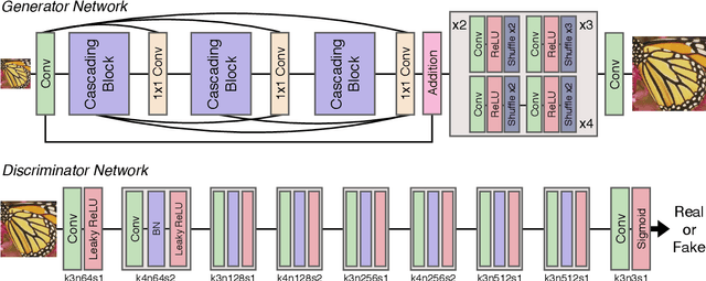
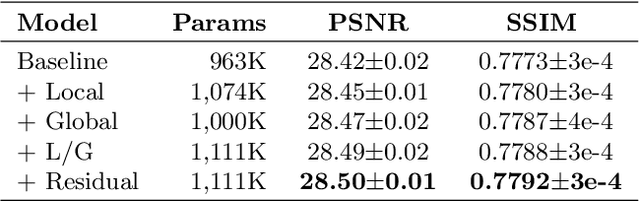
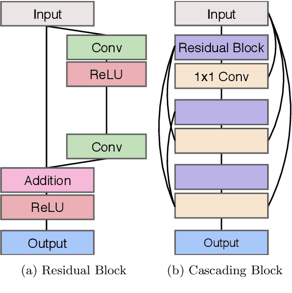
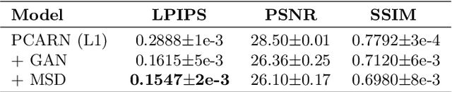
Recent progress in the deep learning-based models has improved single-image super-resolution significantly. However, despite their powerful performance, many models are difficult to apply to the real-world applications because of the heavy computational requirements. To facilitate the use of a deep learning model in such demands, we focus on keeping the model fast and lightweight while maintaining its accuracy. In detail, we design an architecture that implements a cascading mechanism on a residual network to boost the performance with limited resources via multi-level feature fusion. Moreover, we adopt group convolution and weight-tying for our proposed model in order to achieve extreme efficiency. In addition to the traditional super-resolution task, we apply our methods to the photo-realistic super-resolution field using the adversarial learning paradigm and a multi-scale discriminator approach. By doing so, we show that the performances of the proposed models surpass those of the recent methods, which have a complexity similar to ours, for both traditional pixel-based and perception-based tasks.
Adversarial Learning of Semantic Relevance in Text to Image Synthesis
Dec 12, 2018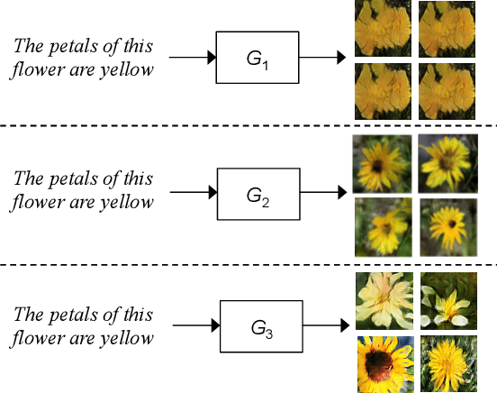
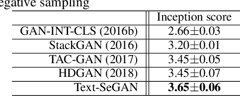
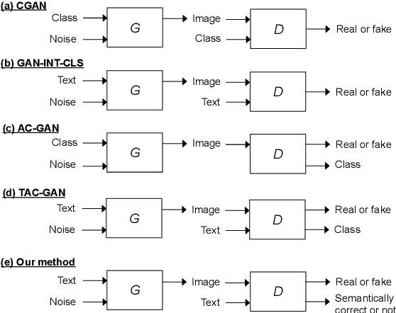
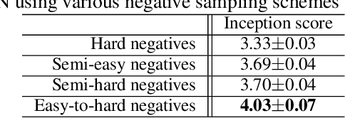
We describe a new approach that improves the training of generative adversarial nets (GANs) for synthesizing diverse images from a text input. Our approach is based on the conditional version of GANs and expands on previous work leveraging an auxiliary task in the discriminator. Our generated images are not limited to certain classes and do not suffer from mode collapse while semantically matching the text input. A key to our training methods is how to form positive and negative training examples with respect to the class label of a given image. Instead of selecting random training examples, we perform negative sampling based on the semantic distance from a positive example in the class. We evaluate our approach using the Oxford-102 flower dataset, adopting the inception score and multi-scale structural similarity index (MS-SSIM) metrics to assess discriminability and diversity of the generated images. The empirical results indicate greater diversity in the generated images, especially when we gradually select more negative training examples closer to a positive example in the semantic space.
A smartphone based multi input workflow for non-invasive estimation of haemoglobin levels using machine learning techniques
Nov 29, 2020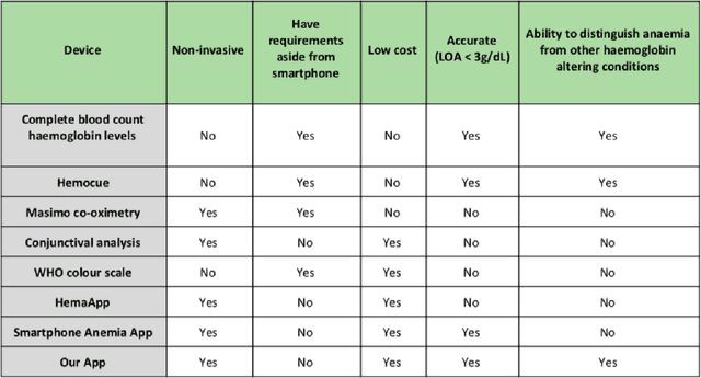
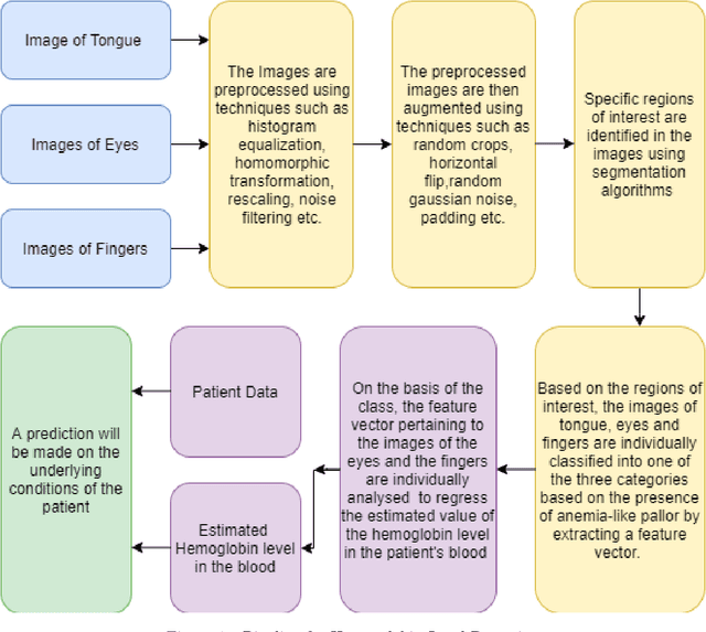
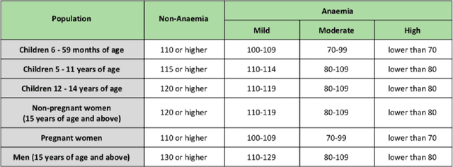
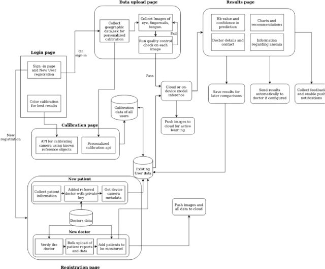
We suggest a low cost, non invasive healthcare system that measures haemoglobin levels in patients and can be used as a preliminary diagnostic test for anaemia. A combination of image processing, machine learning and deep learning techniques are employed to develop predictive models to measure haemoglobin levels. This is achieved through the color analysis of the fingernail beds, palpebral conjunctiva and tongue of the patients. This predictive model is then encapsulated in a healthcare application. This application expedites data collection and facilitates active learning of the model. It also incorporates personalized calibration of the model for each patient, assisting in the continual monitoring of the haemoglobin levels of the patient. Upon validating this framework using data, it can serve as a highly accurate preliminary diagnostic test for anaemia.
W-Net: Dense Semantic Segmentation of Subcutaneous Tissue in Ultrasound Images by Expanding U-Net to Incorporate Ultrasound RF Waveform Data
Aug 27, 2020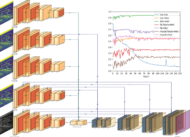
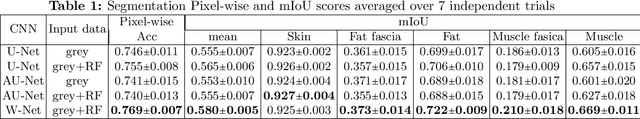
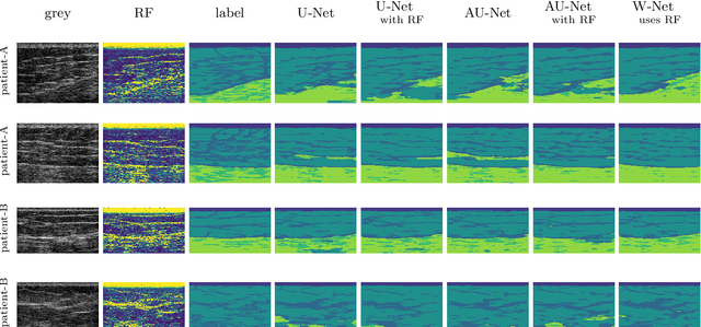
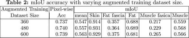
We present W-Net, a novel Convolution Neural Network (CNN) framework that employs raw ultrasound waveforms from each A-scan, typically referred to as ultrasound Radio Frequency (RF) data, in addition to the gray ultrasound image to semantically segment and label tissues. Unlike prior work, we seek to label every pixel in the image, without the use of a background class. To the best of our knowledge, this is also the first deep-learning or CNN approach for segmentation that analyses ultrasound raw RF data along with the gray image. International patent(s) pending [PCT/US20/37519]. We chose subcutaneous tissue (SubQ) segmentation as our initial clinical goal since it has diverse intermixed tissues, is challenging to segment, and is an underrepresented research area. SubQ potential applications include plastic surgery, adipose stem-cell harvesting, lymphatic monitoring, and possibly detection/treatment of certain types of tumors. A custom dataset consisting of hand-labeled images by an expert clinician and trainees are used for the experimentation, currently labeled into the following categories: skin, fat, fat fascia/stroma, muscle and muscle fascia. We compared our results with U-Net and Attention U-Net. Our novel \emph{W-Net}'s RF-Waveform input and architecture increased mIoU accuracy (averaged across all tissue classes) by 4.5\% and 4.9\% compared to regular U-Net and Attention U-Net, respectively. We present analysis as to why the Muscle fascia and Fat fascia/stroma are the most difficult tissues to label. Muscle fascia in particular, the most difficult anatomic class to recognize for both humans and AI algorithms, saw mIoU improvements of 13\% and 16\% from our W-Net vs U-Net and Attention U-Net respectively.
Spatiotemporal tomography based on scattered multiangular signals and its application for resolving evolving clouds using moving platforms
Dec 06, 2020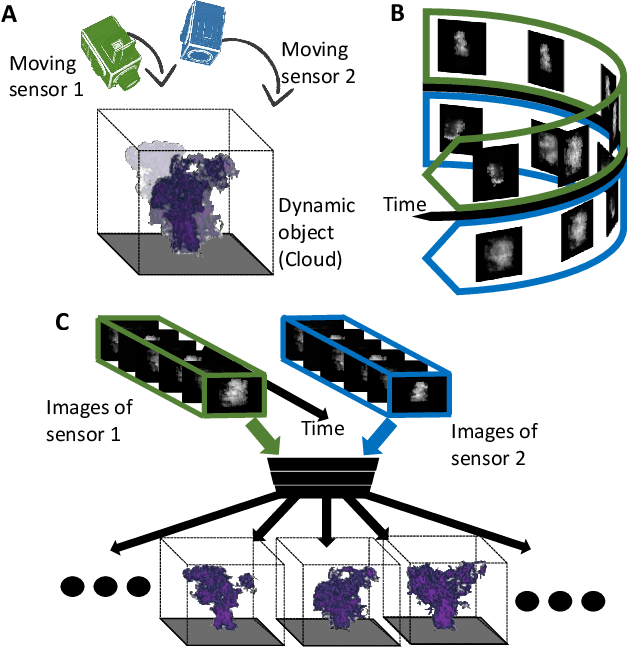
We derive computed tomography (CT) of a time-varying volumetric translucent object, using a small number of moving cameras. We particularly focus on passive scattering tomography, which is a non-linear problem. We demonstrate the approach on dynamic clouds, as clouds have a major effect on Earth's climate. State of the art scattering CT assumes a static object. Existing 4D CT methods rely on a linear image formation model and often on significant priors. In this paper, the angular and temporal sampling rates needed for a proper recovery are discussed. If these rates are used, the paper leads to a representation of the time-varying object, which simplifies 4D CT tomography. The task is achieved using gradient-based optimization. We demonstrate this in physics-based simulations and in an experiment that had yielded real-world data.
Joint Learning of 3D Shape Retrieval and Deformation
Jan 19, 2021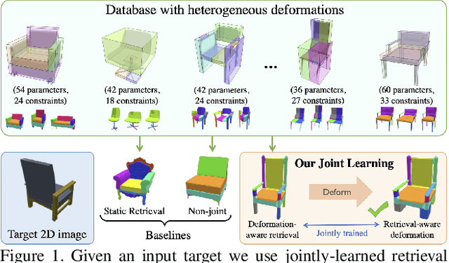
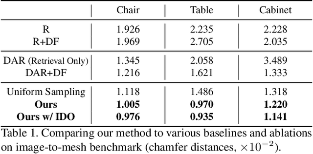
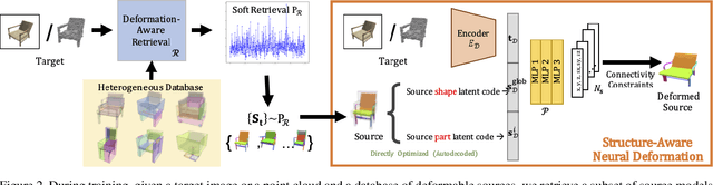
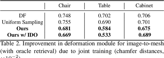
We propose a novel technique for producing high-quality 3D models that match a given target object image or scan. Our method is based on retrieving an existing shape from a database of 3D models and then deforming its parts to match the target shape. Unlike previous approaches that independently focus on either shape retrieval or deformation, we propose a joint learning procedure that simultaneously trains the neural deformation module along with the embedding space used by the retrieval module. This enables our network to learn a deformation-aware embedding space, so that retrieved models are more amenable to match the target after an appropriate deformation. In fact, we use the embedding space to guide the shape pairs used to train the deformation module, so that it invests its capacity in learning deformations between meaningful shape pairs. Furthermore, our novel part-aware deformation module can work with inconsistent and diverse part-structures on the source shapes. We demonstrate the benefits of our joint training not only on our novel framework, but also on other state-of-the-art neural deformation modules proposed in recent years. Lastly, we also show that our jointly-trained method outperforms a two-step deformation-aware retrieval that uses direct optimization instead of neural deformation or a pre-trained deformation module.