Hip and pelvic fractures are serious injuries with life-threatening complications. However, diagnostic errors of fractures in pelvic X-rays (PXRs) are very common, driving the demand for computer-aided diagnosis (CAD) solutions. A major challenge lies in the fact that fractures are localized patterns that require localized analyses. Unfortunately, the PXRs residing in hospital picture archiving and communication system do not typically specify region of interests. In this paper, we propose a two-stage hip and pelvic fracture detection method that executes localized fracture classification using weakly supervised ROI mining. The first stage uses a large capacity fully-convolutional network, i.e., deep with high levels of abstraction, in a multiple instance learning setting to automatically mine probable true positive and definite hard negative ROIs from the whole PXR in the training data. The second stage trains a smaller capacity model, i.e., shallower and more generalizable, with the mined ROIs to perform localized analyses to classify fractures. During inference, our method detects hip and pelvic fractures in one pass by chaining the probability outputs of the two stages together. We evaluate our method on 4 410 PXRs, reporting an area under the ROC curve value of 0.975, the highest among state-of-the-art fracture detection methods. Moreover, we show that our two-stage approach can perform comparably to human physicians (even outperforming emergency physicians and surgeons), in a preliminary reader study of 23 readers.
Automatic parsing of anatomical objects in X-ray images is critical to many clinical applications in particular towards image-guided invention and workflow automation. Existing deep network models require a large amount of labeled data. However, obtaining accurate pixel-wise labeling in X-ray images relies heavily on skilled clinicians due to the large overlaps of anatomy and the complex texture patterns. On the other hand, organs in 3D CT scans preserve clearer structures as well as sharper boundaries and thus can be easily delineated. In this paper, we propose a novel model framework for learning automatic X-ray image parsing from labeled CT scans. Specifically, a Dense Image-to-Image network (DI2I) for multi-organ segmentation is first trained on X-ray like Digitally Reconstructed Radiographs (DRRs) rendered from 3D CT volumes. Then we introduce a Task Driven Generative Adversarial Network (TD-GAN) architecture to achieve simultaneous style transfer and parsing for unseen real X-ray images. TD-GAN consists of a modified cycle-GAN substructure for pixel-to-pixel translation between DRRs and X-ray images and an added module leveraging the pre-trained DI2I to enforce segmentation consistency. The TD-GAN framework is general and can be easily adapted to other learning tasks. In the numerical experiments, we validate the proposed model on 815 DRRs and 153 topograms. While the vanilla DI2I without any adaptation fails completely on segmenting the topograms, the proposed model does not require any topogram labels and is able to provide a promising average dice of 85% which achieves the same level accuracy of supervised training (88%).
2D/3D image registration to align a 3D volume and 2D X-ray images is a challenging problem due to its ill-posed nature and various artifacts presented in 2D X-ray images. In this paper, we propose a multi-agent system with an auto attention mechanism for robust and efficient 2D/3D image registration. Specifically, an individual agent is trained with dilated Fully Convolutional Network (FCN) to perform registration in a Markov Decision Process (MDP) by observing a local region, and the final action is then taken based on the proposals from multiple agents and weighted by their corresponding confidence levels. The contributions of this paper are threefold. First, we formulate 2D/3D registration as a MDP with observations, actions, and rewards properly defined with respect to X-ray imaging systems. Second, to handle various artifacts in 2D X-ray images, multiple local agents are employed efficiently via FCN-based structures, and an auto attention mechanism is proposed to favor the proposals from regions with more reliable visual cues. Third, a dilated FCN-based training mechanism is proposed to significantly reduce the Degree of Freedom in the simulation of registration environment, and drastically improve training efficiency by an order of magnitude compared to standard CNN-based training method. We demonstrate that the proposed method achieves high robustness on both spine cone beam Computed Tomography data with a low signal-to-noise ratio and data from minimally invasive spine surgery where severe image artifacts and occlusions are presented due to metal screws and guide wires, outperforming other state-of-the-art methods (single agent-based and optimization-based) by a large margin.
3-D image registration, which involves aligning two or more images, is a critical step in a variety of medical applications from diagnosis to therapy. Image registration is commonly performed by optimizing an image matching metric as a cost function. However, this task is challenging due to the non-convex nature of the matching metric over the plausible registration parameter space and insufficient approaches for a robust optimization. As a result, current approaches are often customized to a specific problem and sensitive to image quality and artifacts. In this paper, we propose a completely different approach to image registration, inspired by how experts perform the task. We first cast the image registration problem as a "strategy learning" process, where the goal is to find the best sequence of motion actions (e.g. up, down, etc.) that yields image alignment. Within this approach, an artificial agent is learned, modeled using deep convolutional neural networks, with 3D raw image data as the input, and the next optimal action as the output. To cope with the dimensionality of the problem, we propose a greedy supervised approach for an end-to-end training, coupled with attention-driven hierarchical strategy. The resulting registration approach inherently encodes both a data-driven matching metric and an optimal registration strategy (policy). We demonstrate, on two 3-D/3-D medical image registration examples with drastically different nature of challenges, that the artificial agent outperforms several state-of-art registration methods by a large margin in terms of both accuracy and robustness.
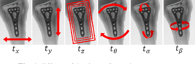
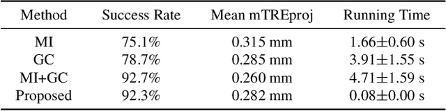
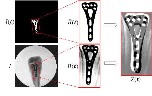
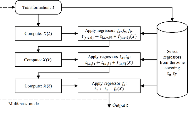
In this paper, we present a Convolutional Neural Network (CNN) regression approach for real-time 2-D/3-D registration. Different from optimization-based methods, which iteratively optimize the transformation parameters over a scalar-valued metric function representing the quality of the registration, the proposed method exploits the information embedded in the appearances of the Digitally Reconstructed Radiograph and X-ray images, and employs CNN regressors to directly estimate the transformation parameters. The CNN regressors are trained for local zones and applied in a hierarchical manner to break down the complex regression task into simpler sub-tasks that can be learned separately. Our experiment results demonstrate the advantage of the proposed method in computational efficiency with negligible degradation of registration accuracy compared to intensity-based methods.